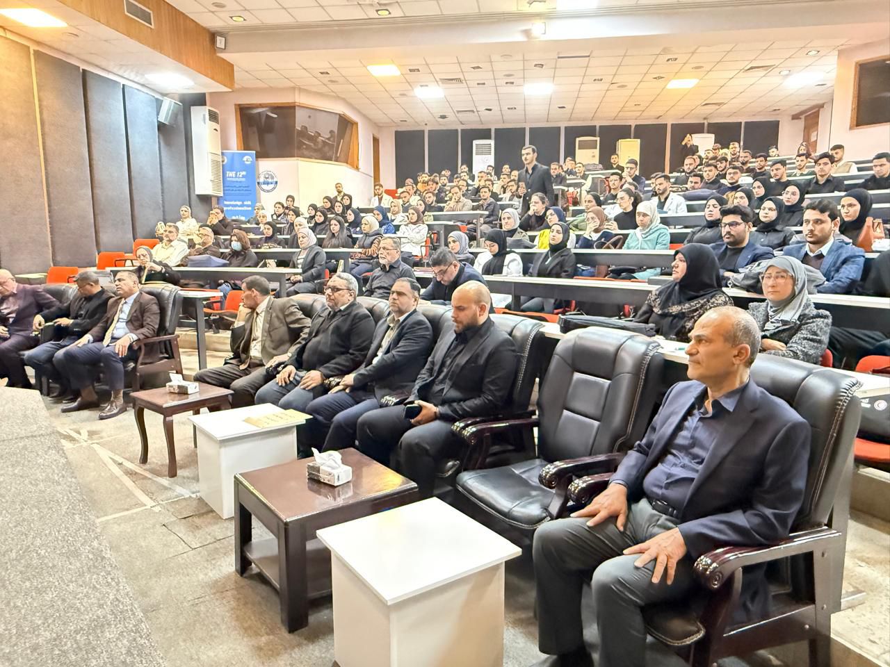
تتشرف كلية الطب – جامعة النهرين بالإعلان عن إقامة المؤتمر العلمي الدولي الثالث عشر لجامعة النهرين تحت عنوان الشراكة الأكاديمية، التميز البحثي، والتعاون العالمي في الطب يهدف المؤتمر إلى جمع الباحثين والأكاديميين والأطباء والتربويين من داخل العراق وخارجه، لتعزيز البحث العلمي، ودعم الشراكات الأكاديمية، وتبادل الخبرات العلمية في مختلف المجالات الطبية
جامعة النهرين .. بين الواقع والمستقبل تسعى جامعة النهرين الى تأمين قاعدة بيانات من الخبرات العلمية تتوافر فيها قابليات الابداع وتطبيق الجودة والسعي للحصول على الاعتمادية الدولية, كما تسعى
تسعى لجان الاعتمادية البرامجية في الكلية للحصول على الاعتماد الدولي من خلال تبني برنامج تعليمي مواكب للشروط والمواصفات المحددة من قبل الفدرالية العالمية للتعليم الطبي (WFME) ومواصلة عملية التطويرمن خلال التقييم الذاتي المستمر، وقد حصلت كلية طب النهرين في نيسان 2024 على الاعتماد الدولي من خلال حصولها على اعتماد هيئة مؤسسات التعليم العالي ونظام الجودة الاردنية (AQACHEI) المعتمدة من قبل (WFME). وكانت الكلية قد حصلت عام 2022 على إعتماد مشروط من قبل المجلس الوطني لاعتماد كليات الطب في العراق (NCAMC) وتعمل على الحصول على الاعتماد الكامل من نفس المجلس ومن خلال عمل ثلاث لجان للاعتمادية وهي:
اللجنة التوجيهية برئاسة السيد عميد الكلية
اللجنة الرئيسية برئاسة السيد معاون العميد للشؤون العلمية والطلبة
اللجان الفرعية للاعتمادية
وخلال سنوات عمل لجان الاعتمادية في الكلية منذ عام 2007 تمت كتابة اربعة طبعات من التقرير الذاتي للكلية للاعوام
2010 - 2014 - 2018 (وتعديله عام 2021) - 2023
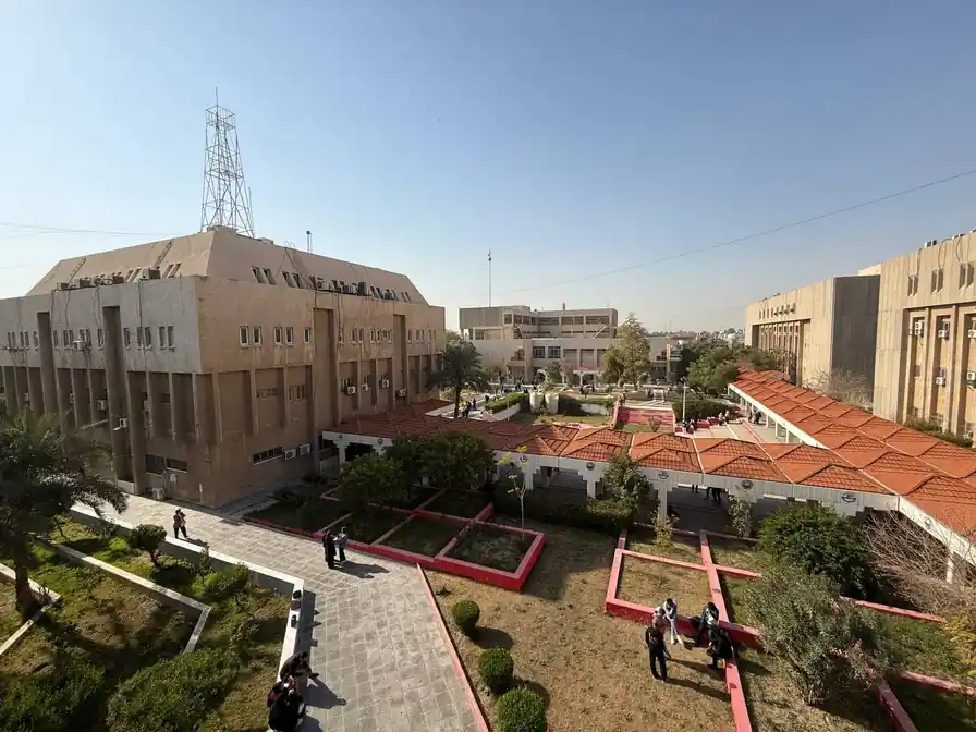
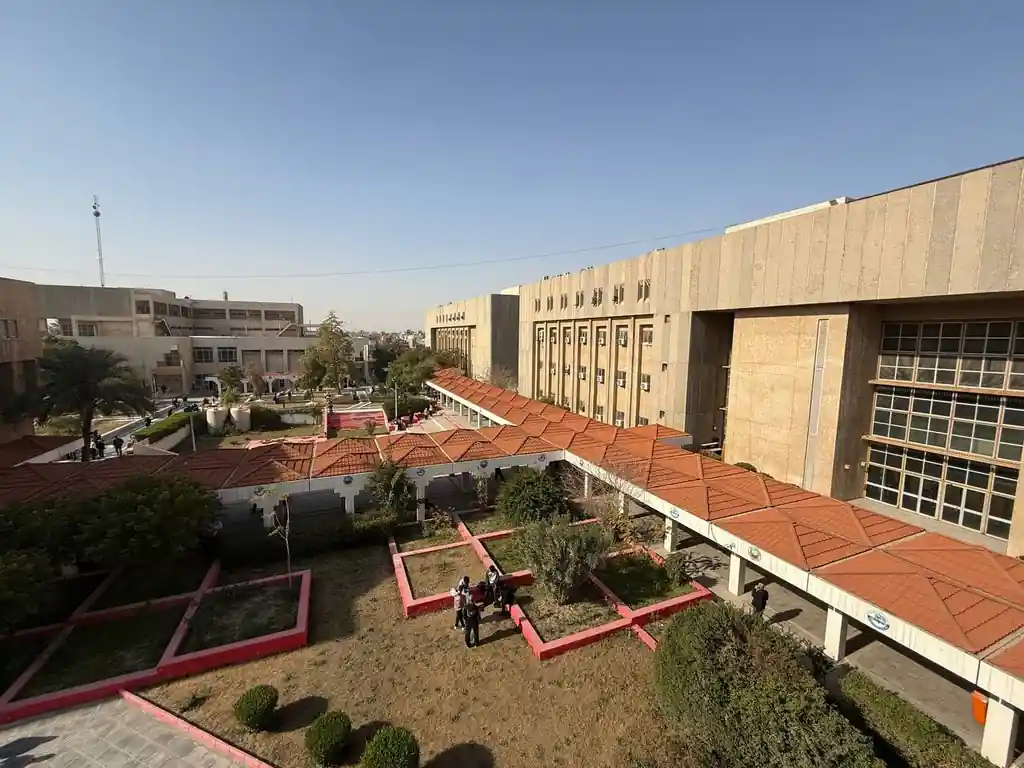
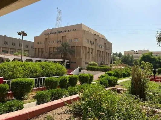
كلية الطب - جامعة النهرين
كلية الطب جامعة النهرين تناقش رسالة ماجستير حول توظيف المتراكبات النانوية لتعزيز فعالية علاج سرطان الثدي
أجرى فرع علم وظائف الأعضاء/ الفيزياء الطبية في كلية الطب جامعة النهرين في يوم الخميس الموافق 2026/3/5 مناقشة طالبة الماجستير غدير هيثم مصطفى الزاملي ع...
ندوة علمية في كلية الطب بجامعة النهرين حول التشخيص المتكامل لعقد الغدة الدرقية
برعاية السيد عميد كلية الطب جامعة النهرين الاستاذ الدكتور انيس خليل نايل نظم فرع الطب الباطني بالتعاون مع فرع الجراحة في الكلية وبمشاركة المجلس العراق...
كلية الطب في جامعة النهرين تحتضن مناقشات بحوث طلبة البورد العراقي في اختصاص الجراحة العامة
احتضنت القاعات الدراسية في كلية الطب بجامعة النهرين مناقشات عدد من بحوث طلبة البورد العراقي في اختصاص الجراحة العامة ضمن متطلبات برنامج التدريب التخصص...
...
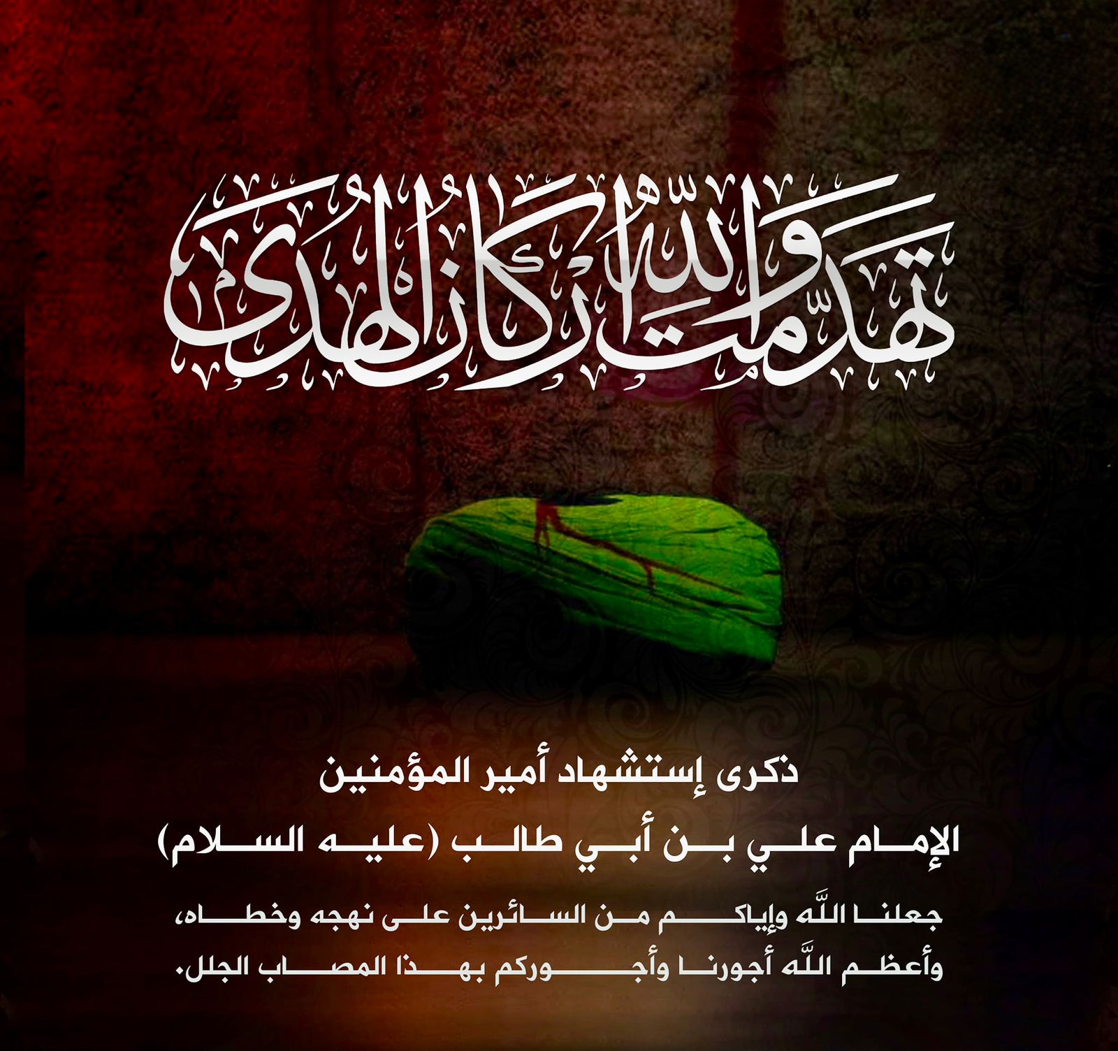
مناسبة ذكرى استشهاد امير المؤمنين علي بن ابي طالب (ع) .
بمناسبة ذكرى استشهاد امير المؤمنين علي بن ابي طالب عليه السلام اتقدم بخالص التعازي والمواساة الى الامة الاسلامية والى ابناء شعبنا العزيز والى اساتذة...
مجلس تأبيني لطالبة المرحلة الرابعة براء علاء هادي
أقيم في كلية الطب جامعة النهرين مجلس تأبيني على روح طالبة المرحلة الرابعة ( براء علاء هادي ) وذلك يوم الثلاثاء الموافق 3/3 في قاعة المؤتمرات بالبناية...
مبادرة رمضانية لتعزيز التكافل الاجتماعي في كلية الطب جامعة النهرين
برعاية السيد عميد كلية الطب في جامعة النهرين الاستاذ الدكتور انيس خليل نايل وانسجاما مع الاهداف النبيلة والانسانية لشهر رمضان المبارك نظمت وحدة شؤون ا...
...
كلية الطب جامعة النهرين تناقش رسالة ماجستير حول توظيف المتراكبات النانوية لتعزيز فعالية علاج سرطان الثدي
أجرى فرع علم وظائف الأعضاء/ الفيزياء الطبية في كلية الطب جامعة النهرين في يوم الخميس الموافق 2026/3/5 مناقشة طالبة الماجستير غدير هيثم مصطفى الزاملي ع...
ندوة علمية في كلية الطب بجامعة النهرين حول التشخيص المتكامل لعقد الغدة الدرقية
برعاية السيد عميد كلية الطب جامعة النهرين الاستاذ الدكتور انيس خليل نايل نظم فرع الطب الباطني بالتعاون مع فرع الجراحة في الكلية وبمشاركة المجلس العراق...
كلية الطب في جامعة النهرين تحتضن مناقشات بحوث طلبة البورد العراقي في اختصاص الجراحة العامة
احتضنت القاعات الدراسية في كلية الطب بجامعة النهرين مناقشات عدد من بحوث طلبة البورد العراقي في اختصاص الجراحة العامة ضمن متطلبات برنامج التدريب التخصص...
...
دورة حول التقييم السريري ضمن برنامج الامتحانات والتقويم في كلية الطب بجامعة النهرين
برعاية السيد عميد كلية الطب جامعة النهرين الاستاذ الدكتور ( انيس خليل نايل ) تستمر فعاليات دورة الامتحانات والتقويم التي تنظمها الوحدة المختصة في الكل...
المؤتمر العلمي الدولي الثالث عشر لكلية الطب - جامعة النهرين
المؤتمر العلمي الدولي الثالث عشر لكلية الطب - جامعة النهرين
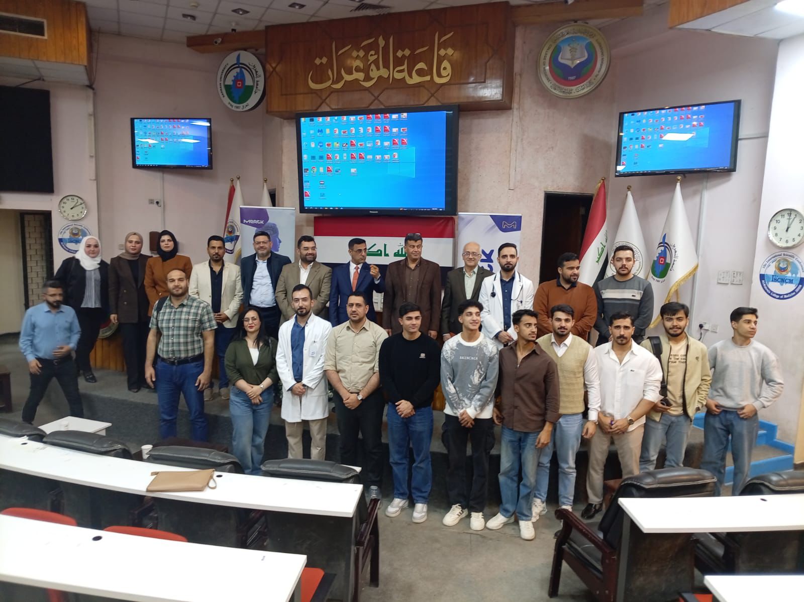
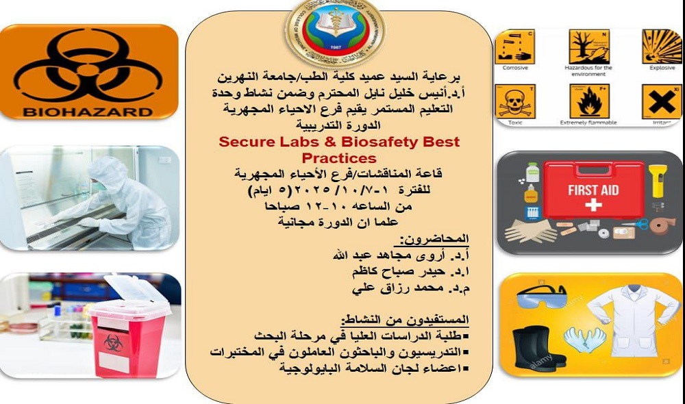
دورة تدريبية يقيمها فرع الاحياء المجهرية حول المختبرات الآمنة وأفضل ممارسات السلامة البيولوجية
المختبرات الآمنة وأفضل ممارسات السلامة البيولوجيةالمحاضرون:الفئة المستهدفة:طلبة الدراسات العليا في مرحلة البحثالتدريسيون والموظفون العاملون في المختبر...
...
ترقية الى مرتبة الأستاذية
تتويجاً للجهود العلمية والبحثية المبذولة من قبل الأستاذ المساعد الدكتور ( قحطان عدنان مهدي ) عضو الكادر التدريسي في فرع الجراحة اختصاص (طب وجراحة عا...
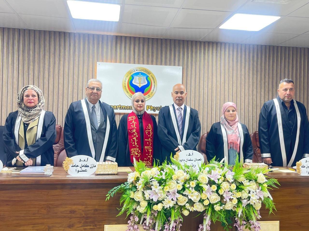
ترقية المدرس الدكتورة نسرين احمد ناصرالى مرتبة استاذ مساعد
تتويجاً للجهود العلمية والبحثية المبذولة من قبل المدرس الدكتورة نسرين احمد ناصر / التدريسية في فرع الكيمياء والكيمياء الحياتية اختصاص ( علوم كيميا...
ترقية الأستاذ المساعد الدكتورة اقبال غالب فرهود الى مرتبة أستاذ
تتويجاً للجهود العلمية والبحثية المبذولة من قبل الأستاذ المساعد الدكتورة اقبال غالب فرهود التدريسية في فرع الطب الباطني اختصاص (طب وجراحة عامة - الأمر...
...
اعلان
برعاية عمادة كلية الطب في جامعة النهرين وبالتعاون مع فرع الجراحة ممثلا برئيس الفرع الاستاذ الدكتور بشار عباس وعضو الهيئة التدريسية الاستاذ المساعد ا...
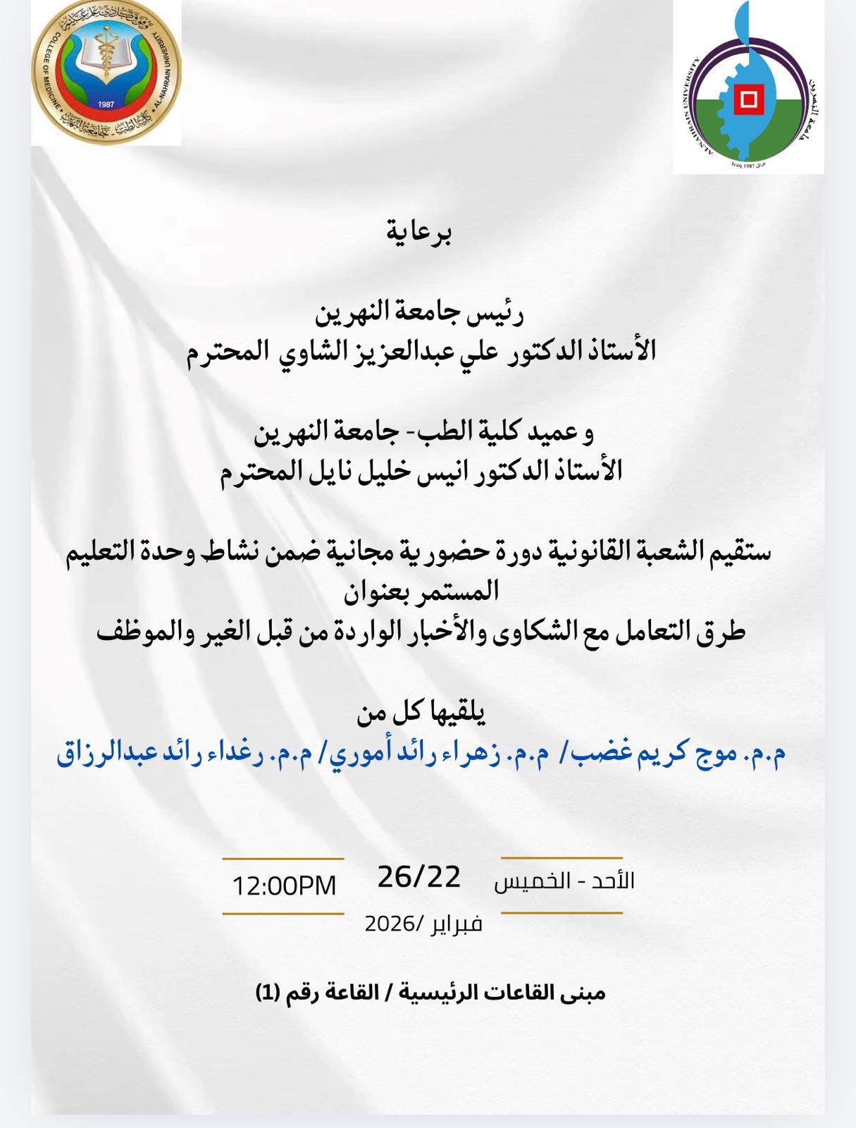
ستُقيم الشعبة القانونية في كلية الطب – جامعة النهرين دورة تدريبية حول طرق التعامل مع الشكاوى والإخبار الواردة من قبل الغير والموظفين
.
برعاية السيد رئيس جامعة النهرين الأستاذ الدكتور علي عبدالعزيز الشاوي،والسيد عميد كلية الطب – جامعة النهرين الأستاذ الدكتور أنيس خليل نايل، تُقيم الو...
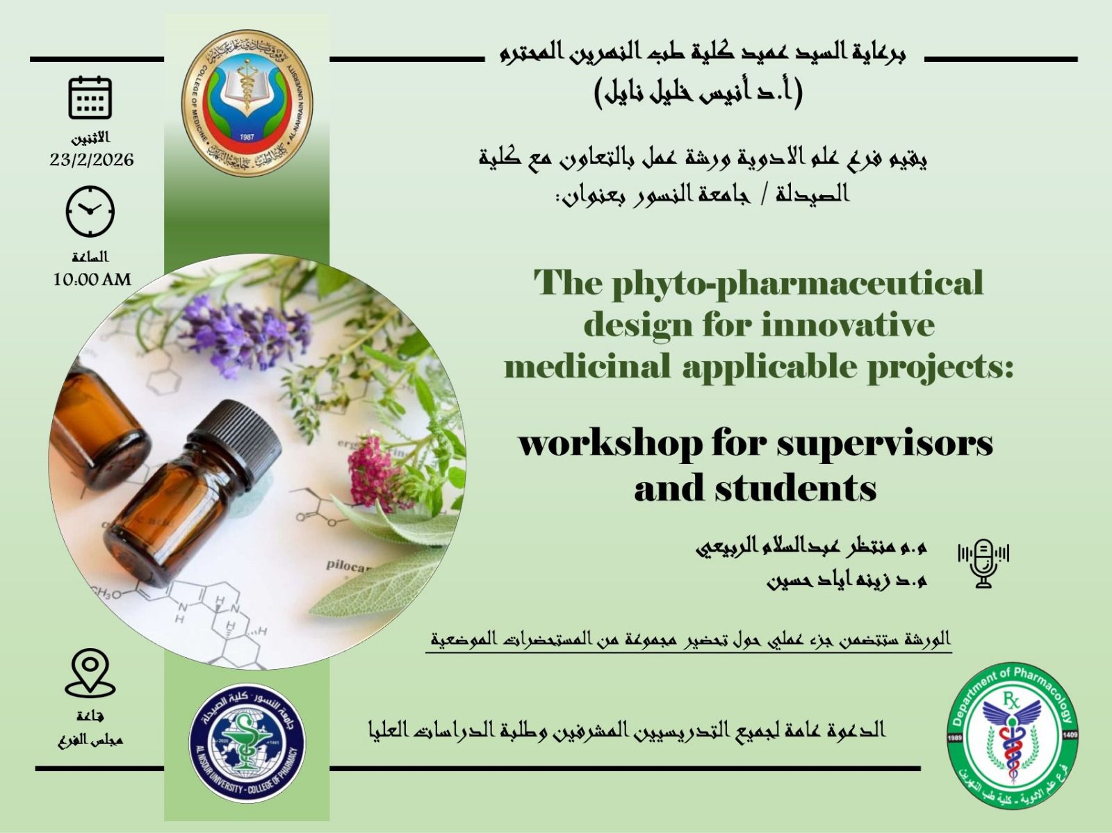
تصميم المستحضرات الصيدلانية النباتية لمشاريع طبية مبتكرة قابلة للتطبيق
...
المنشورات العلمية للاساتذة
LATE-ONSET THYROID EYE DISEASE IN A PATIENT WITH MULTIPLE COMORBIDITIES: A CASE REPORT
المؤلفون: Zaid Qahtan Abd Al Razq and Mahmood Shakir Khudhair
تاريخ النشر:
Assessment of Immunohistochemical Expression to the TP63 Protein in Premalignant and Malignant Disor...
المؤلفون: Jasim Mohammed Muhsin, Azhar A. F. Al-Attraqchi, Ban J. Qasim
تاريخ النشر:
Diagnostic Performance of Urinary Antigen Test and Molecular Method for Detection of Legionella pneu...
المؤلفون: Thanaa R Abdulrahman (1) , Shaymaa A Gauad (2) , Amar K Muhamad (3) , Jabbar S Hassan (1)
تاريخ النشر:
Broad-spectrum bioactivities of a sulfated heteropolysaccharide from Bacillus tequilensis MYG163: an...
المؤلفون: Maha Abdullah Alwaili,Mohammed A. Alshehrib,Thanaa R. Abdulrahman,Faisal Miqad K. Albaqami,Abdullah Alghamdi,Mona Othman I. Albureikan,Taghreed Shamrani,Abdullah Albelasi,Mohammad El-Nablaway,Samy Selim &Ahmed GhareebORCID Icon show less
تاريخ النشر:
Interstitial Lung Disease and Pulmonary Arterial Hypertension Screening Practices in Systemic Sclero...
المؤلفون: Rajaie Namas1 ,Sarah Al Qassimi1 , Jawahir Alameri1
, Samar Al Emadi2 , Nelly Ziade3 , Ahlam Almarzooqi4 ,
Farida Al Balushi5
, Mohammed A. Omair6 , Taha Qardaghi7 , Yasameen Abbas Humadi8 , Mohamed Alawlaqi1
,
Hanan Al Rayes9 , Saadeya Naji10, Fatima Haji11, Fajer Altamimi12, Mansour Alazmi13 , Hani Shatnawi14, Waleed Hafiz15 ,
Deena Ahmed16, Mariam Almansoori16, Jamal Al Saleh17 , Hazem Rifaai18, Mahdi Abusalameh18 , Zaki Abou Zahr19,
Hiba Khogali20, Sehriban Diab16, Maha Anbar21, Adeeba Al Herz22 , Amal Elganzoury23, Shaima Ewila24, Basant Elnady25 ,
Wafa Madanat26, Imad Uthman27 , Asia Mubashir1 , Mohamed Elarabi1 , Suzan Attar
تاريخ النشر:
Iraqi Registry Data Proves Safety and Efficacy of Switching to Adalimumab Biosimilar in Treating Rh...
المؤلفون: Yasameen Abbas Humadi1*
, Nabaa Ihsan2
, Asal Adnan3
, Marwa Moayad4
, Ali AlKazzaz5
, Avin Maroof6
, Ali
Abdulrahman7
, Nazar Abdulateef3
, Mohammad AlOsami3
, Faiq Gorial3
, Mohammed Altahhan2
, Zahraa Almansi2
, Ali
AlNoori2
تاريخ النشر:
Decoding eQTLs: unraveling their role in spondyloarthropathies pathogenesis and potential therapeuti...
المؤلفون: Nabaa Ihsan Awadh 1
*, Asal Adnan Ridha1
,
Marwah Muayad Younus 2
, Yasameen Abbas Humadi 3
,
Faiq I. Gorial 1
, Khalid Burhan Khalid 4 and Yousif Alaa Rasoo
تاريخ النشر:
Molecular and Serological Detection of Human Parvovirus B19 in a Sample of Iraqi Patients with Acute...
المؤلفون: Mohammed A. Rahema, Arwa M. Al-Shuwaikh, Waseem F. Al Tameemi
تاريخ النشر:
Detection of Human Parvovirus B19 IgG in a Group of Iraqi Children with Newly onset Type 1 Diabetes...
المؤلفون: Ealaf A. Khudair , Arwa M. Al-Shuwaikh, Dawood S. Abdoun
تاريخ النشر:
Investigation of CTLA-4 gene polymorphisms in asample of Iraqi children with newly diagnosed type 1...
المؤلفون: Ealaf Abbas Khudair, Arwa Mujahid Al-Shuwaikh, Dawood Salman Abdoun
تاريخ النشر:
The Association of Reactive Oxygen Species with High-Risk Human Papilloma Virus Infection and Some S...
المؤلفون: Mustafa A. Dawood, Arwa M. Al-Shuwaikh, Ula M. Al-Kawaz
تاريخ النشر:
From Virome to Biomarker: Insights into the Role of Torque Teno Virus in Health and Ecosystems