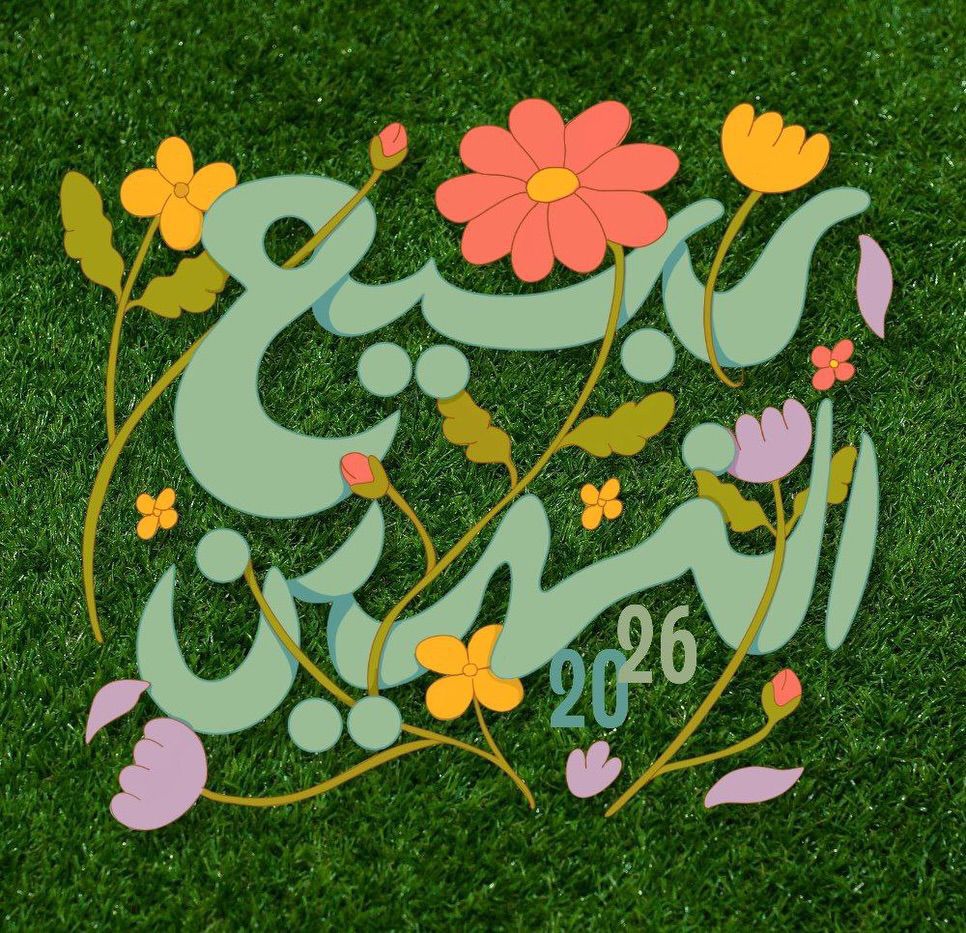
مهرجان ربيع النهرين
تتشرف كلية الطب / جامعة النهرين بدعوتكم لحضور مهرجان ربيع النهرين 🌿📅 الاثنين 16 شباط 2026📍 في أروقة وحدائق الكليةيتضمن المهرجان حفل داخل القاعة ا...
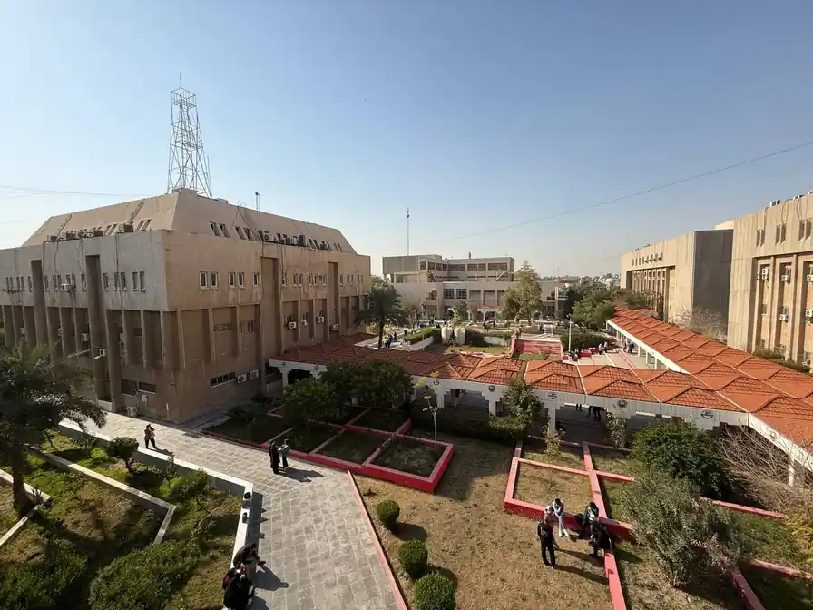
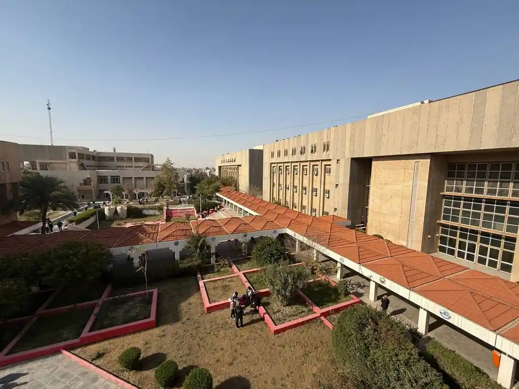
قسم عن الكلية يحتوي على المعلومات والتفاصيل

رؤية الكلية، رسالتها وأهدافها الاست...

قائمة عمداء الكلية الحاليين والسابق...

أعضاء مجلس الكلية والطاقم الإداري

خريجي الكلية والمخرجات التعليمية

البحوث والمنشورات العلمية لأعضاء هي...

المناهج الأكاديمية والبرامج الدراسي...

قوانين وتشريعات ووثائق تنظيمية للكل...

خدمات وخبرات المركز الاستشاري للكلي...


مبادرات وبرامج التنمية المستدامة في...
تتشرف كلية الطب – جامعة النهرين بالإعلان عن إقامة المؤتمر العلمي الدولي الثالث عشر لجامعة النهرين تحت عنوان الشراكة الأكاديمية، التميز البحثي، والتعاون العالمي في الطب يهدف المؤتمر إلى جمع الباحثين والأكاديميين والأطباء والتربويين من داخل العراق وخارجه، لتعزيز البحث العلمي، ودعم الشراكات الأكاديمية، وتبادل الخبرات العلمية في مختلف المجالات الطبية
جامعة النهرين .. بين الواقع والمستقبل تسعى جامعة النهرين الى تأمين قاعدة بيانات من الخبرات العلمية تتوافر فيها قابليات الابداع وتطبيق الجودة والسعي للحصول على الاعتمادية الدولية, كما تسعى
تسعى لجان الاعتمادية البرامجية في الكلية للحصول على الاعتماد الدولي من خلال تبني برنامج تعليمي مواكب للشروط والمواصفات المحددة من قبل الفدرالية العالمية للتعليم الطبي (WFME) ومواصلة عملية التطويرمن خلال التقييم الذاتي المستمر، وقد حصلت كلية طب النهرين في نيسان 2024 على الاعتماد الدولي من خلال حصولها على اعتماد هيئة مؤسسات التعليم العالي ونظام الجودة الاردنية (AQACHEI) المعتمدة من قبل (WFME). وكانت الكلية قد حصلت عام 2022 على إعتماد مشروط من قبل المجلس الوطني لاعتماد كليات الطب في العراق (NCAMC) وتعمل على الحصول على الاعتماد الكامل من نفس المجلس ومن خلال عمل ثلاث لجان للاعتمادية وهي:
وخلال سنوات عمل لجان الاعتمادية في الكلية منذ عام 2007 تمت كتابة اربعة طبعات من التقرير الذاتي للكلية للاعوام
2010 - 2014 - 2018 (وتعديله عام 2021) - 2023
تتشرف كلية الطب / جامعة النهرين بدعوتكم لحضور مهرجان ربيع النهرين 🌿📅 الاثنين 16 شباط 2026📍 في أروقة وحدائق الكليةيتضمن المهرجان حفل داخل القاعة ا...
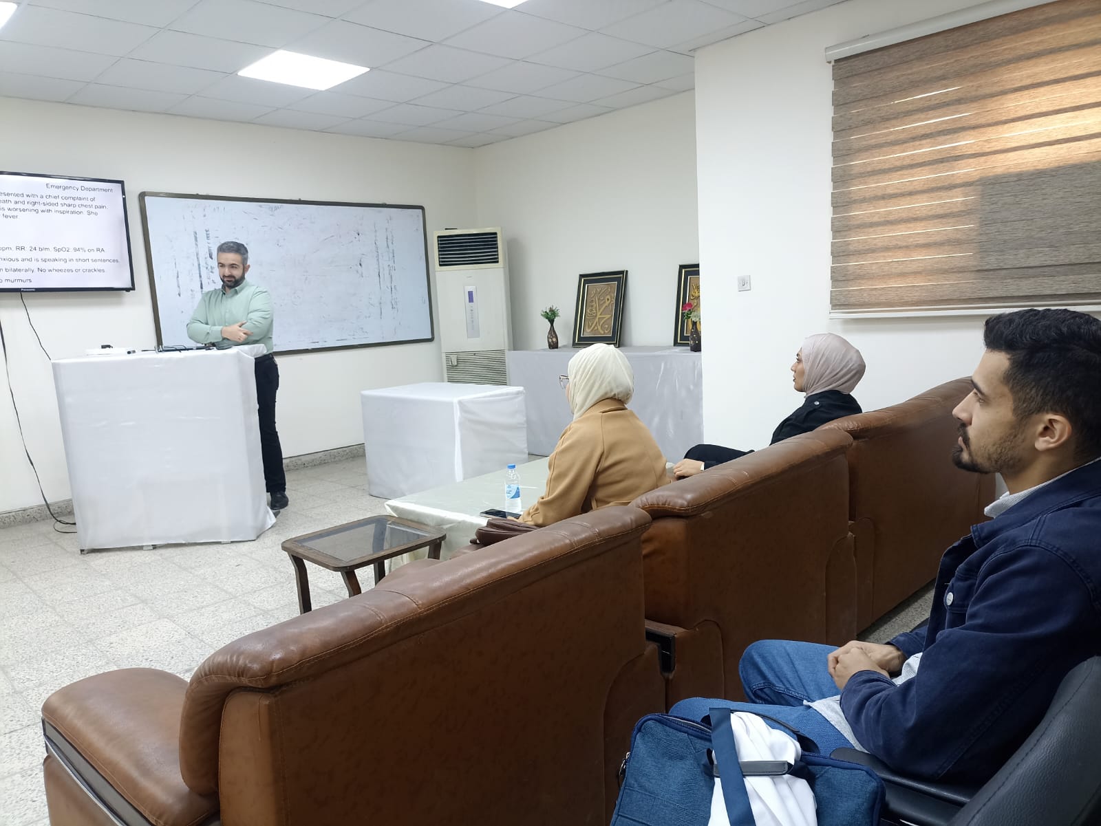
ضمن برنامج المحاضرات والتدريب والاشراف على طلبة البورد العراقي للاختصاصات الطبية اختصاص الكيمياء المرضية القيت يوم الاربعاء الموافق 2026 2 11 محاضرة ع...
اجريت مناقشات طلبة المجلس العراقي اختصاص علم الامراض/امراض الدم للطالبتين نور سعد مفلح/ اشراف الأستاذ المساعد الدكتور بسام محمد حميد عنوان البحث الم...
تتشرف كلية الطب / جامعة النهرين بدعوتكم لحضور مهرجان ربيع النهرين 🌿📅 الاثنين 16 شباط 2026📍 في أروقة وحدائق الكليةيتضمن المهرجان حفل داخل القاعة ا...
برعاية السيد عميد كلية الطب جامعة النهرين الاستاذ الدكتور ( انيس خليل نايل ) وضمن نشاطات فرع الجراحة وبالتعاون مع كلية الجراحيين الامريكية اقامت كلية...
الأخوات والأخوة منتسبو كلية الطب في جامعة النهرين المحترمونتحية طيبة…في الوقت الذي نُثمّن ونُقدّر عاليًا الجهود المبذولة من قبل كافة كوادرنا المخلصة،...
ضمن برنامج المحاضرات والتدريب والاشراف على طلبة البورد العراقي للاختصاصات الطبية اختصاص الكيمياء المرضية القيت يوم الاربعاء الموافق 2026 2 11 محاضرة ع...
اجريت مناقشات طلبة المجلس العراقي اختصاص علم الامراض/امراض الدم للطالبتين نور سعد مفلح/ اشراف الأستاذ المساعد الدكتور بسام محمد حميد عنوان البحث الم...
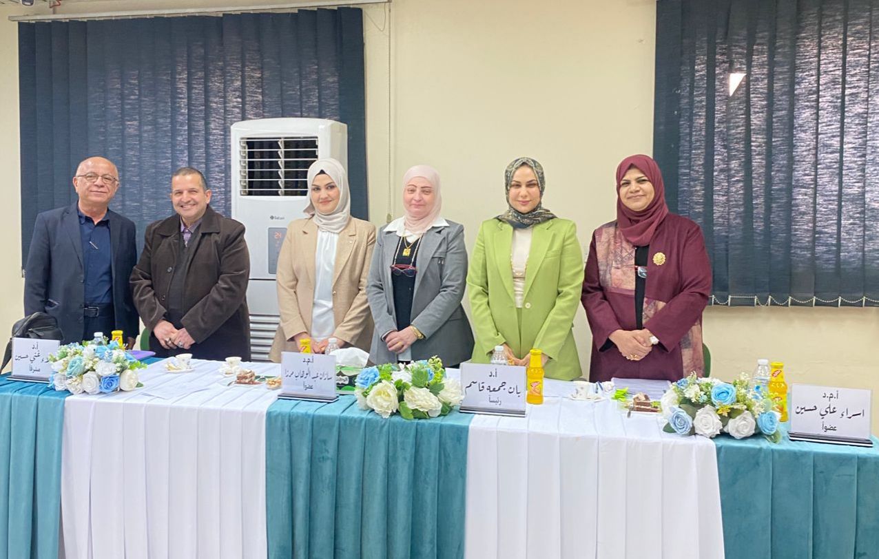
تم بتاريخ ٢٠٢٦/٢/٩ الساعة التاسعة صباحا في فرع علم الامراض والطب العدلي مناقشة بحث طالبة المجلس العراقي للاختصاصات الطبية ( هبة محمد سلمان) (التقييم...

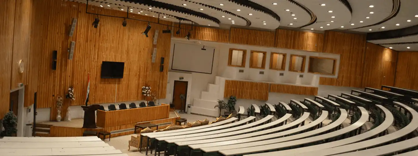
المؤتمر العلمي الدولي الثالث عشر لكلية الطب - جامعة النهرين
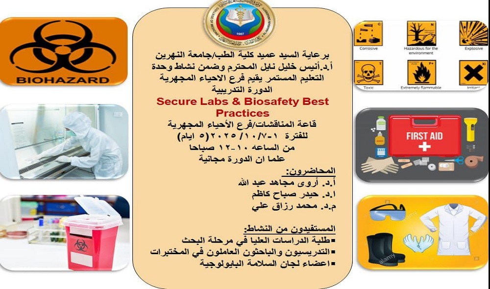
المختبرات الآمنة وأفضل ممارسات السلامة البيولوجيةالمحاضرون:الفئة المستهدفة:طلبة الدراسات العليا في مرحلة البحثالتدريسيون والموظفون العاملون في المختبر...

تتويجاً للجهود العلمية والبحثية المبذولة من قبل المدرس الدكتورة نسرين احمد ناصر / التدريسية في فرع الكيمياء والكيمياء الحياتية اختصاص ( علوم كيميا...
تتويجاً للجهود العلمية والبحثية المبذولة من قبل الأستاذ المساعد الدكتورة اقبال غالب فرهود التدريسية في فرع الطب الباطني اختصاص (طب وجراحة عامة - الأمر...
تتويجاً للجهود العلمية والبحثية المبذولة من قبل الأستاذ المساعد الدكتورة اقبال غالب فرهود التدريسية في فرع الطب الباطني اختصاص (طب وجراحة عامة - الأمر...
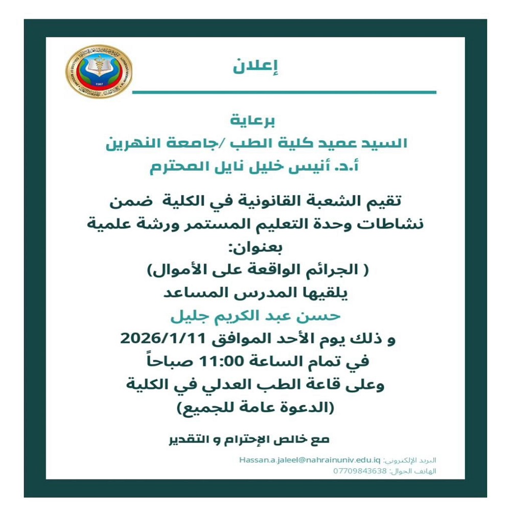
برعاية السيد عميد كلية الطب / جامعة النهرين الأستاذ الدكتور أنيس خليل نايل المحترم، تقيم الشعبة القانونية في الكلية ضمن نشاطات وحدة التعليم المستمر ور...
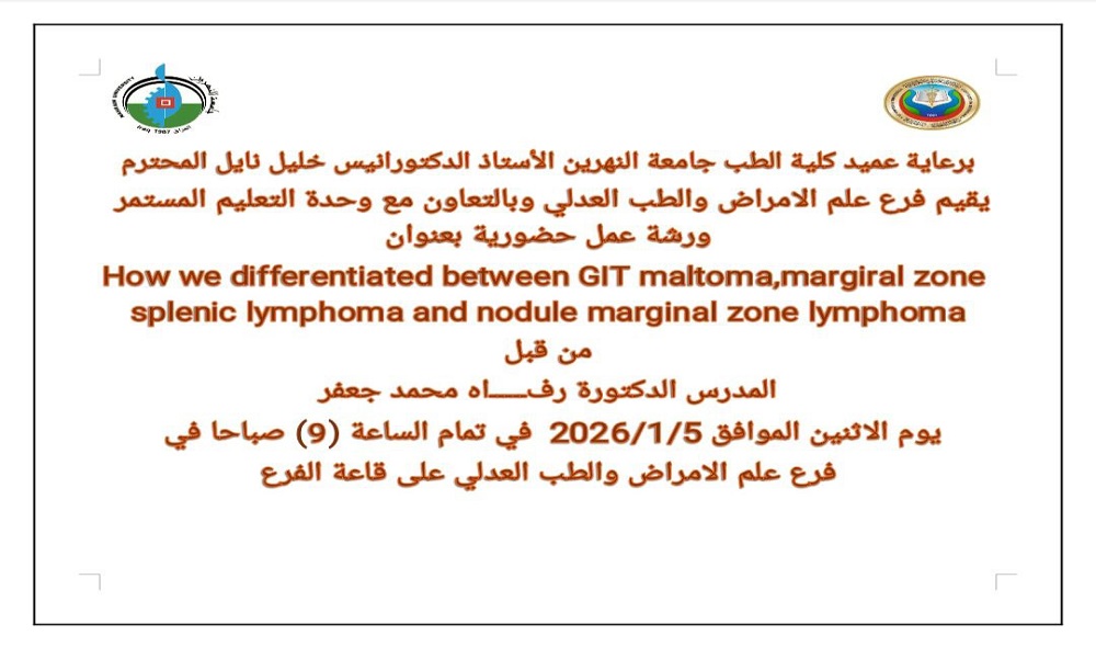
برعاية عميد كلية الطب في جامعة النهرين الأستاذ الدكتور خليل نايل، يقيم فرع علم الأمراض والطب العدلي وبالتعاون مع وحدة التعليم المستمر ورشة عمل حضورية...