
تهنئة
تتقدم عمادة كلية الطب جامعة النهرين والمتمثلة بالسيد عميد الكلية الاستاذ الدكتور انيس خليل نايل بأسمى آيات التهاني والتبريكات الى أبناء شعبنا العزيز و...
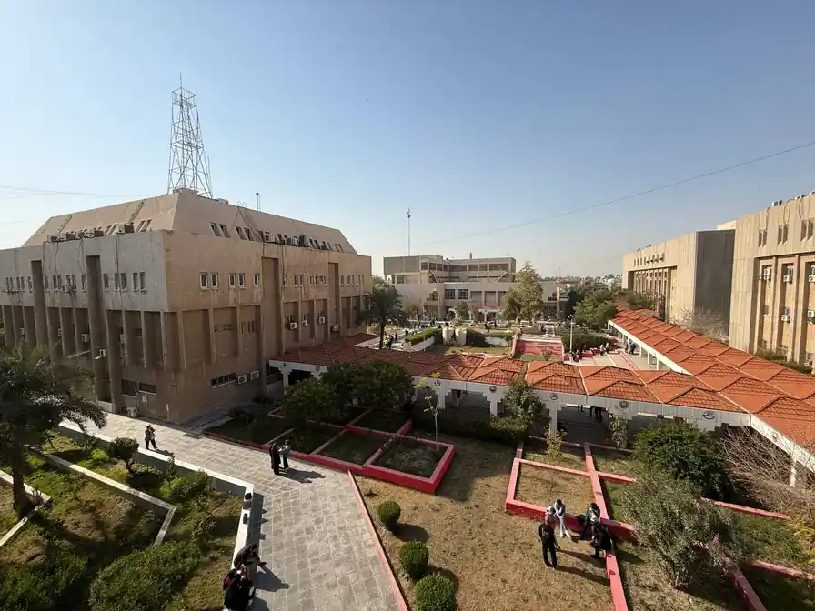
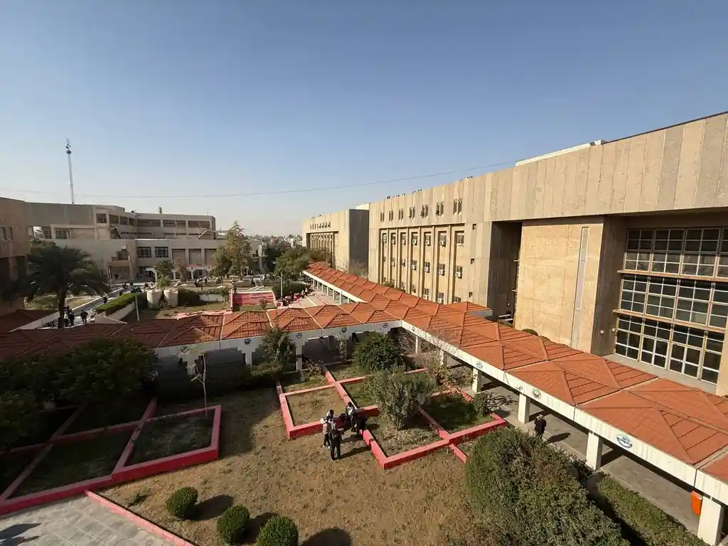
قسم عن الكلية يحتوي على المعلومات والتفاصيل

رؤية الكلية، رسالتها وأهدافها الاست...

قائمة عمداء الكلية الحاليين والسابق...

أعضاء مجلس الكلية والطاقم الإداري

خريجي الكلية والمخرجات التعليمية

البحوث والمنشورات العلمية لأعضاء هي...

المناهج الأكاديمية والبرامج الدراسي...

قوانين وتشريعات ووثائق تنظيمية للكل...

خدمات وخبرات المركز الاستشاري للكلي...


مبادرات وبرامج التنمية المستدامة في...
تتشرف كلية الطب – جامعة النهرين بالإعلان عن إقامة المؤتمر العلمي الدولي الثالث عشر لجامعة النهرين تحت عنوان الشراكة الأكاديمية، التميز البحثي، والتعاون العالمي في الطب يهدف المؤتمر إلى جمع الباحثين والأكاديميين والأطباء والتربويين من داخل العراق وخارجه، لتعزيز البحث العلمي، ودعم الشراكات الأكاديمية، وتبادل الخبرات العلمية في مختلف المجالات الطبية
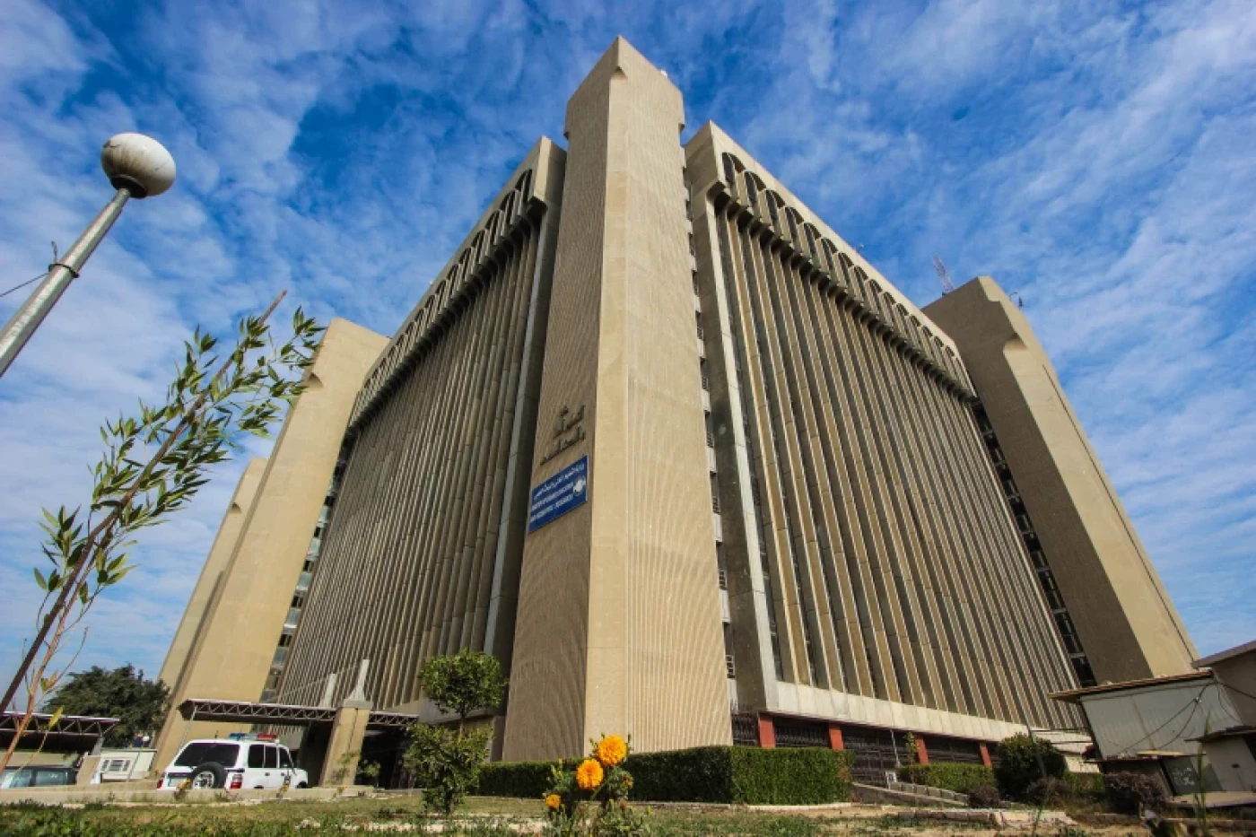
جامعة النهرين .. بين الواقع والمستقبل تسعى جامعة النهرين الى تأمين قاعدة بيانات من الخبرات العلمية تتوافر فيها قابليات الابداع وتطبيق الجودة والسعي للحصول على الاعتمادية الدولية, كما تسعى
تسعى لجان الاعتمادية البرامجية في الكلية للحصول على الاعتماد الدولي من خلال تبني برنامج تعليمي مواكب للشروط والمواصفات المحددة من قبل الفدرالية العالمية للتعليم الطبي (WFME) ومواصلة عملية التطويرمن خلال التقييم الذاتي المستمر، وقد حصلت كلية طب النهرين في نيسان 2024 على الاعتماد الدولي من خلال حصولها على اعتماد هيئة مؤسسات التعليم العالي ونظام الجودة الاردنية (AQACHEI) المعتمدة من قبل (WFME). وكانت الكلية قد حصلت عام 2022 على إعتماد مشروط من قبل المجلس الوطني لاعتماد كليات الطب في العراق (NCAMC) وتعمل على الحصول على الاعتماد الكامل من نفس المجلس ومن خلال عمل ثلاث لجان للاعتمادية وهي:
وخلال سنوات عمل لجان الاعتمادية في الكلية منذ عام 2007 تمت كتابة اربعة طبعات من التقرير الذاتي للكلية للاعوام
2010 - 2014 - 2018 (وتعديله عام 2021) - 2023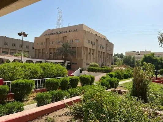
تتقدم عمادة كلية الطب جامعة النهرين والمتمثلة بالسيد عميد الكلية الاستاذ الدكتور انيس خليل نايل بأسمى آيات التهاني والتبريكات الى أبناء شعبنا العزيز و...
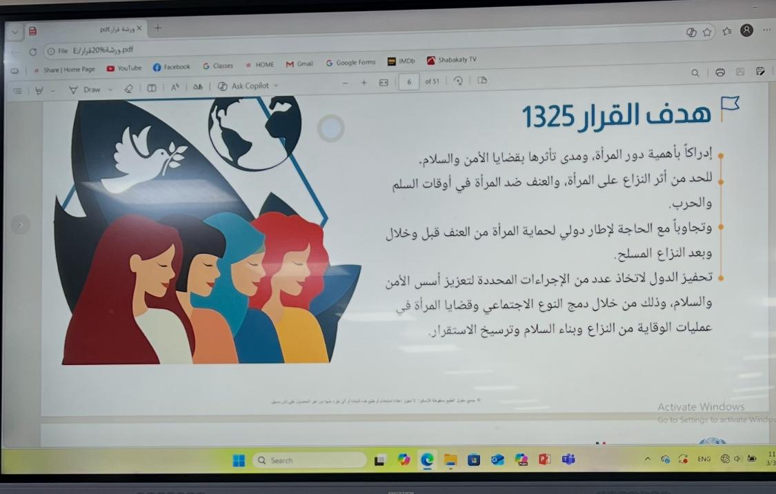
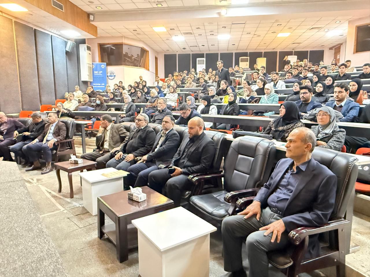
برعاية السيد عميد كلية الطب جامعة النهرين الأستاذ الدكتور ( أنيس خليل نايل ) أقامت وحدة شؤون المرأة في الكلية ندوة علمية بعنوان قرار مجلس الأمن الدولي...
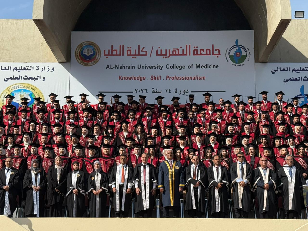
اقامت كلية الطب جامعة النهرين بتاريخ 5 /2 /2026 حفل التقاط صورة التخرج لطلبة المرحلة السادسة الدورة 34 وسط اجواء مفعمة بالفرح والاعتزاز وبرعاية وحضور...
تتقدم عمادة كلية الطب جامعة النهرين والمتمثلة بالسيد عميد الكلية الاستاذ الدكتور انيس خليل نايل بأسمى آيات التهاني والتبريكات الى أبناء شعبنا العزيز و...
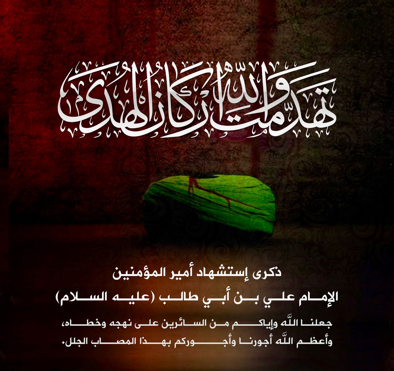
بمناسبة ذكرى استشهاد امير المؤمنين علي بن ابي طالب عليه السلام اتقدم بخالص التعازي والمواساة الى الامة الاسلامية والى ابناء شعبنا العزيز والى اساتذة...
أقيم في كلية الطب جامعة النهرين مجلس تأبيني على روح طالبة المرحلة الرابعة ( براء علاء هادي ) وذلك يوم الثلاثاء الموافق 3/3 في قاعة المؤتمرات بالبناية...
برعاية السيد عميد كلية الطب جامعة النهرين الأستاذ الدكتور ( أنيس خليل نايل ) أقامت وحدة شؤون المرأة في الكلية ندوة علمية بعنوان قرار مجلس الأمن الدولي...
في إنجاز علمي مميز حصلت الدكتورة ( استبرق عبد الرسول الواسطي ) رئيس فرع الكيمياء والكيمياء الحياتية في كليتنا وبالتعاون مع مؤسسات أكاديمية وصحية وطنية...
كرم فرع التشريح البشري في كلية الطب جامعة النهرين عددا من طلبة المرحلة الثانية الحاصلين على درجة الامتياز في مادة التشريح في الامتحان النهائي للفصل ال...

برعاية السيد عميد كلية الطب جامعة النهرين الاستاذ الدكتور ( انيس خليل نايل ) تستمر فعاليات دورة الامتحانات والتقويم التي تنظمها الوحدة المختصة في الكل...

المؤتمر العلمي الدولي الثالث عشر لكلية الطب - جامعة النهرين
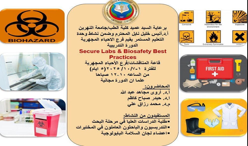
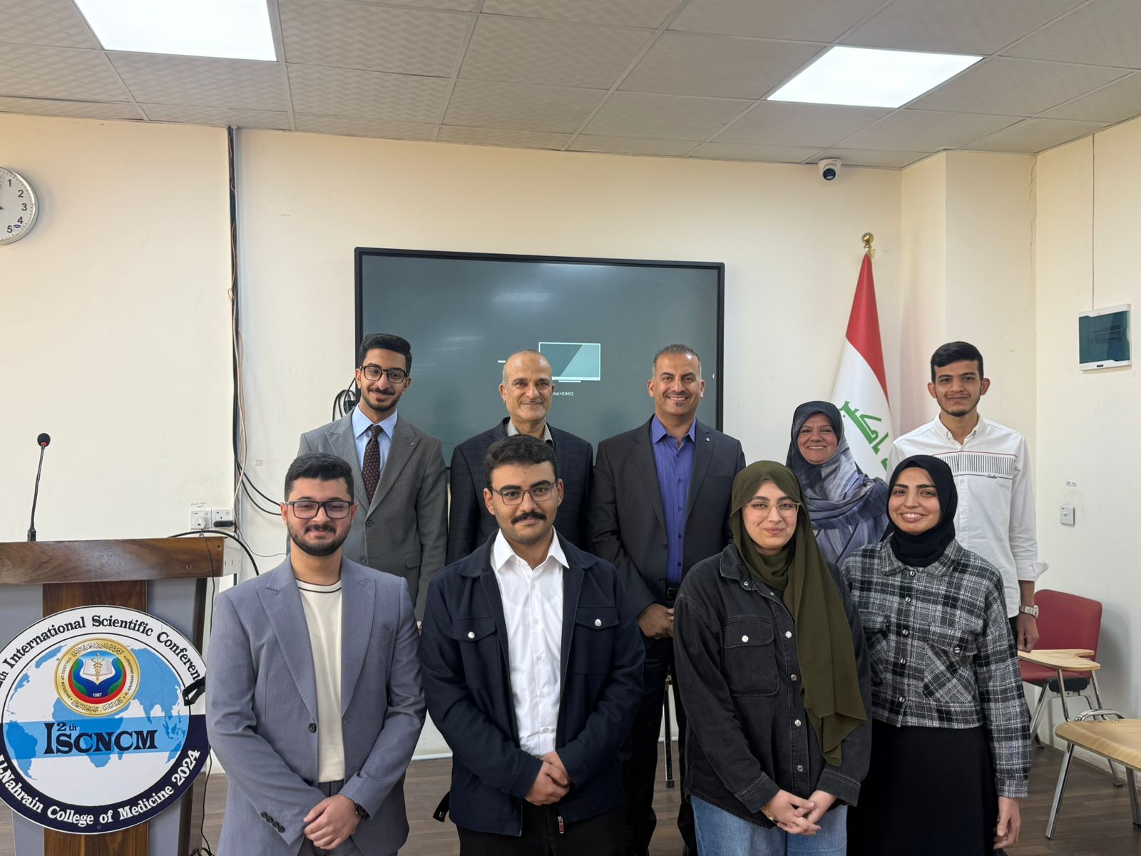
المختبرات الآمنة وأفضل ممارسات السلامة البيولوجيةالمحاضرون:الفئة المستهدفة:طلبة الدراسات العليا في مرحلة البحثالتدريسيون والموظفون العاملون في المختبر...
تتويجاً للجهود العلمية والبحثية المبذولة من قبل الأستاذ المساعد الدكتور ( قحطان عدنان مهدي ) عضو الكادر التدريسي في فرع الجراحة اختصاص (طب وجراحة عا...
تتويجاً للجهود العلمية والبحثية المبذولة من قبل المدرس الدكتورة نسرين احمد ناصر / التدريسية في فرع الكيمياء والكيمياء الحياتية اختصاص ( علوم كيميا...

تتويجاً للجهود العلمية والبحثية المبذولة من قبل الأستاذ المساعد الدكتورة اقبال غالب فرهود التدريسية في فرع الطب الباطني اختصاص (طب وجراحة عامة - الأمر...

نظراً للظروف القاهرة التي تشهدها المنطقة في الوقت الحالي، وحرصاً على ضمان سلامة جميع المشاركين والضيوف، وبسبب تعذر تمكن عدد من الباحثين والمتحدثين و...

برعاية عمادة كلية الطب في جامعة النهرين وبالتعاون مع فرع الجراحة ممثلا برئيس الفرع الاستاذ الدكتور بشار عباس وعضو الهيئة التدريسية الاستاذ المساعد ا...
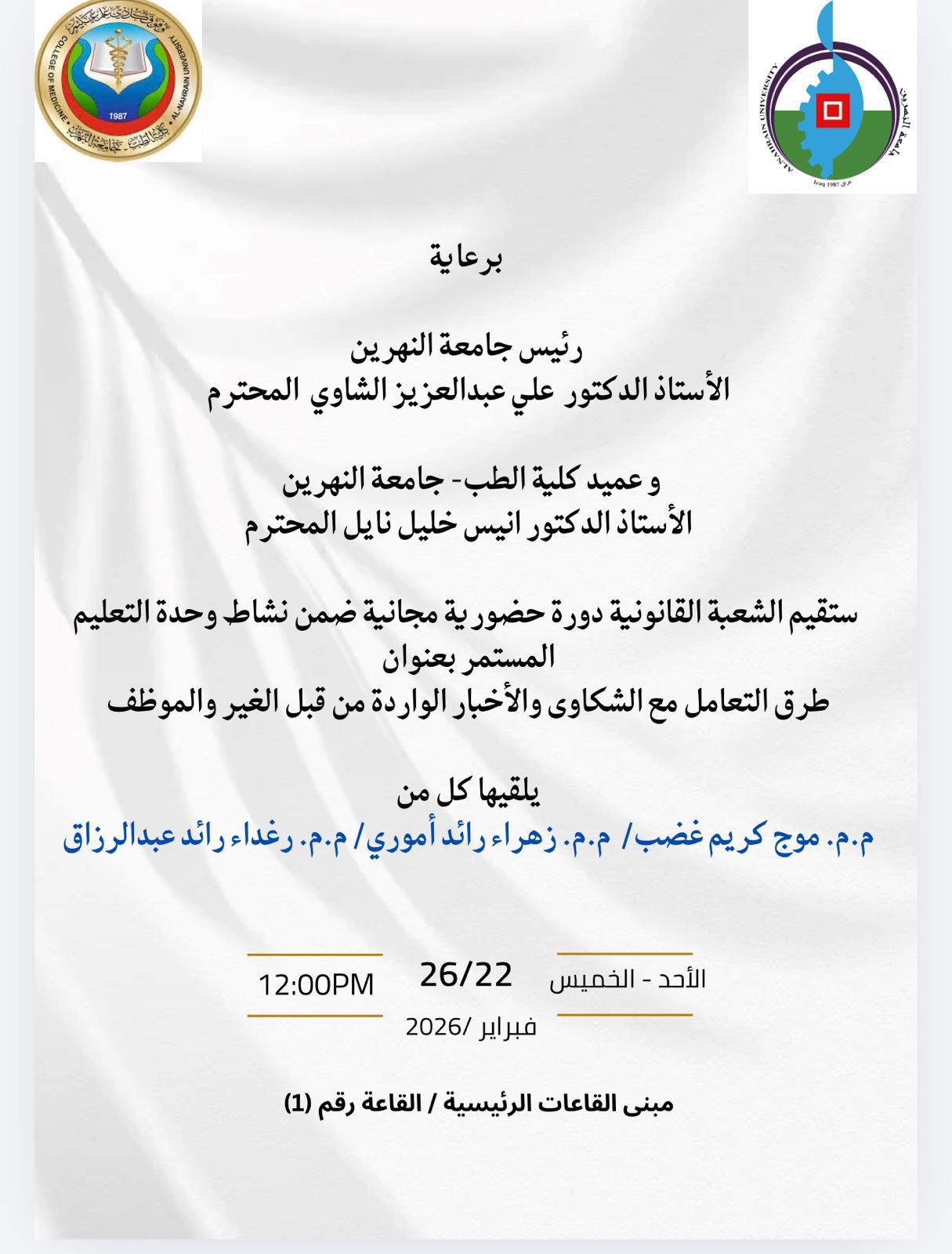
. برعاية السيد رئيس جامعة النهرين الأستاذ الدكتور علي عبدالعزيز الشاوي،والسيد عميد كلية الطب – جامعة النهرين الأستاذ الدكتور أنيس خليل نايل، تُقيم الو...