طلبة المرحلة الثالثة يهدون بوسترات تعليمية لعمادة كلية الطب
تقديرا للجهود المتواصلة التي تبذلها عمادة كلية الطب / جامعة النهرين في دعم المسيرة العلمية والارتقاء بمستوى التدريب السريري للطلبة قام مجموعة من طلبة...
قسم عن الكلية يحتوي على المعلومات والتفاصيل

رؤية الكلية، رسالتها وأهدافها الاست...

قائمة عمداء الكلية الحاليين والسابق...

أعضاء مجلس الكلية والطاقم الإداري

خريجي الكلية والمخرجات التعليمية

تتمثل مهمة مكتبة الكلية في دعم العم...

البحوث والمنشورات العلمية لأعضاء هي...

المناهج الأكاديمية والبرامج الدراسي...

قوانين وتشريعات ووثائق تنظيمية للكل...

خدمات وخبرات المركز الاستشاري للكلي...


مبادرات وبرامج التنمية المستدامة في...
تتشرف كلية الطب – جامعة النهرين بالإعلان عن إقامة المؤتمر العلمي الدولي الثالث عشر لجامعة النهرين تحت عنوان الشراكة الأكاديمية، التميز البحثي، والتعاون العالمي في الطب يهدف المؤتمر إلى جمع الباحثين والأكاديميين والأطباء والتربويين من داخل العراق وخارجه، لتعزيز البحث العلمي، ودعم الشراكات الأكاديمية، وتبادل الخبرات العلمية في مختلف المجالات الطبية
جامعة النهرين .. بين الواقع والمستقبل تسعى جامعة النهرين الى تأمين قاعدة بيانات من الخبرات العلمية تتوافر فيها قابليات الابداع وتطبيق الجودة والسعي للحصول على الاعتمادية الدولية, كما تسعى
تسعى لجان الاعتمادية البرامجية في الكلية للحصول على الاعتماد الدولي من خلال تبني برنامج تعليمي مواكب للشروط والمواصفات المحددة من قبل الفدرالية العالمية للتعليم الطبي (WFME) ومواصلة عملية التطويرمن خلال التقييم الذاتي المستمر، وقد حصلت كلية طب النهرين في نيسان 2024 على الاعتماد الدولي من خلال حصولها على اعتماد هيئة مؤسسات التعليم العالي ونظام الجودة الاردنية (AQACHEI) المعتمدة من قبل (WFME). وكانت الكلية قد حصلت عام 2022 على إعتماد مشروط من قبل المجلس الوطني لاعتماد كليات الطب في العراق (NCAMC) وتعمل على الحصول على الاعتماد الكامل من نفس المجلس ومن خلال عمل ثلاث لجان للاعتمادية وهي:
وخلال سنوات عمل لجان الاعتمادية في الكلية منذ عام 2007 تمت كتابة اربعة طبعات من التقرير الذاتي للكلية للاعوام
2010 - 2014 - 2018 (وتعديله عام 2021) - 2023
تقديرا للجهود المتواصلة التي تبذلها عمادة كلية الطب / جامعة النهرين في دعم المسيرة العلمية والارتقاء بمستوى التدريب السريري للطلبة قام مجموعة من طلبة...
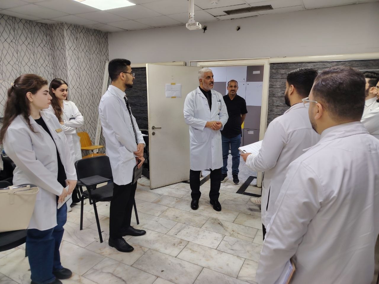
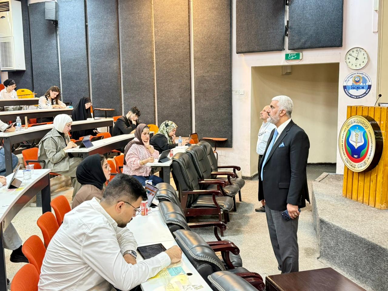
أجرى السيد عميد كلية الطب / جامعة النهرين الأستاذ الدكتور ( أنيس خليل نايل ) جولة تفقدية للاطلاع على سير الامتحانات السريرية النهائية لطلبة المرحلة ال...
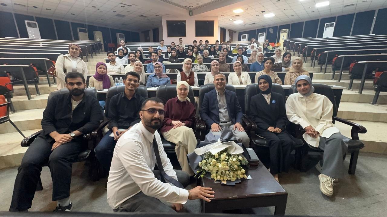
أقامت مجلة أطباء الغد في كلية الطب / جامعة النهرين ورشة تفاعلية متميزة بعنوان : “Humans, Not Machines” بالتعاون مع اختصاصي علاج الأورام والغدد الدكتور...
أجرى السيد عميد كلية الطب / جامعة النهرين الأستاذ الدكتور ( أنيس خليل نايل ) جولة تفقدية للاطلاع على سير الامتحانات السريرية النهائية لطلبة المرحلة ال...
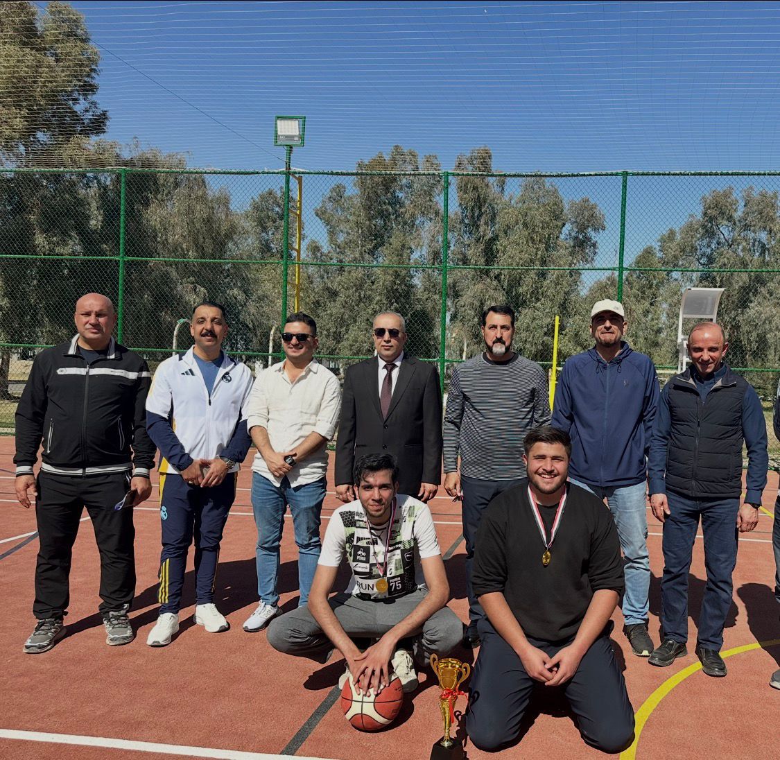
توجت كلية الطب جامعة النهرين بالفوز بالمركز الثاني في بطولة كرة السلة الجامعية التي نظمتها جامعة النهرين ضمن المنهاج السنوي لوزارة التعليم العالي والب...
شهدت كلية الطب / جامعة النهرين انطلاق الامتحانات التقويمية الالكترونية المشتركة لكليات الطب لطلبة المرحلة السادسة للعام الدراسي 2025-2026 وسط اجواء تن...
أقامت مجلة أطباء الغد في كلية الطب / جامعة النهرين ورشة تفاعلية متميزة بعنوان : “Humans, Not Machines” بالتعاون مع اختصاصي علاج الأورام والغدد الدكتور...
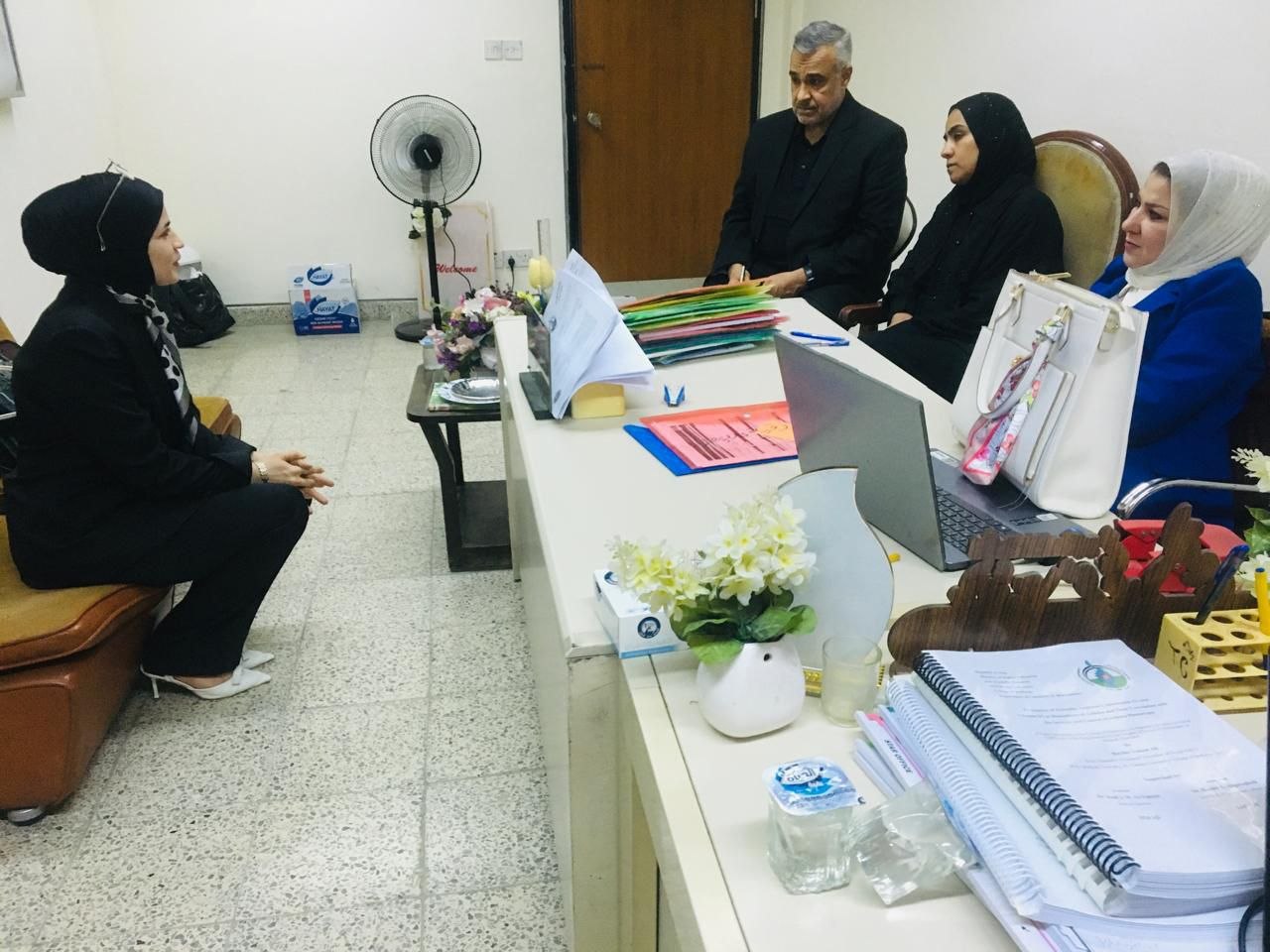
أجرى فرع الكيمياء والكيمياء الحياتية في كلية الطب / جامعة النهرين المقابلات العلمية للمتقدمين إلى برامج الدراسات العليا للعام الدراسي 2026-2027 وذلك ض...
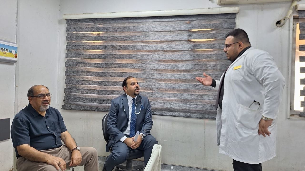
استضاف فرع طب الأطفال في كلية الطب بجامعة النهرين اليوم الأحد الموافق 14/6/2026 نخبة من أساتذة كلية الطب بجامعة الكندي للمشاركة في الامتحان السريري ال...

برعاية السيد عميد كلية الطب جامعة النهرين الاستاذ الدكتور ( انيس خليل نايل ) تستمر فعاليات دورة الامتحانات والتقويم التي تنظمها الوحدة المختصة في الكل...

المؤتمر العلمي الدولي الثالث عشر لكلية الطب - جامعة النهرين
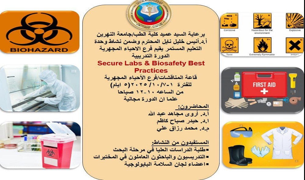
المختبرات الآمنة وأفضل ممارسات السلامة البيولوجيةالمحاضرون:الفئة المستهدفة:طلبة الدراسات العليا في مرحلة البحثالتدريسيون والموظفون العاملون في المختبر...
تتويجاً للجهود العلمية والبحثية المبذولة من قبل المدرس الدكتورة مي فضيل اسطيفان / التدريسية في فرع علم وظائف الاعضاء ( الفسلجة ) اختصاص ( علوم فيزي...
تتويجاً للجهود العلمية والبحثية المبذولة من قبل المدرس الدكتور عبدالرحمن محمدحسن هادي / التدريسي في فرع الاحياء المجهرية اختصاص دقيق ( احياء مجهرية /...
تتويجاً للجهود العلمية والبحثية المبذولة من قبل المدرس الدكتورة نسرين احمد ناصر / التدريسية في فرع الكيمياء والكيمياء الحياتية اختصاص ( علوم كيميا...
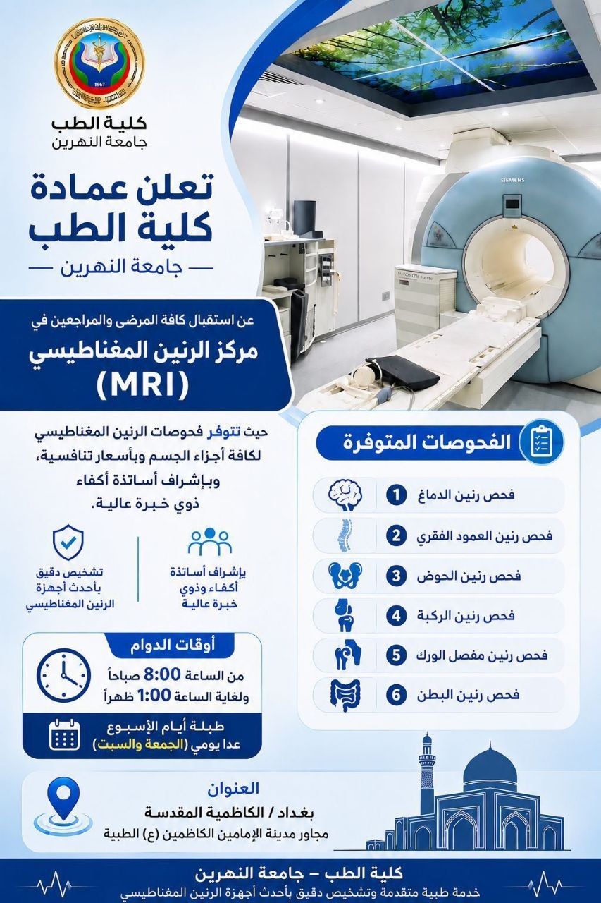
تعلن عمادة كلية الطب – جامعة النهرين عن استقبال كافة المرضى والمراجعين في مركز الرنين المغناطيس حيث تتوفر فحوصات الرنين المغناطيسي لكافة أجزاء الجسم و...
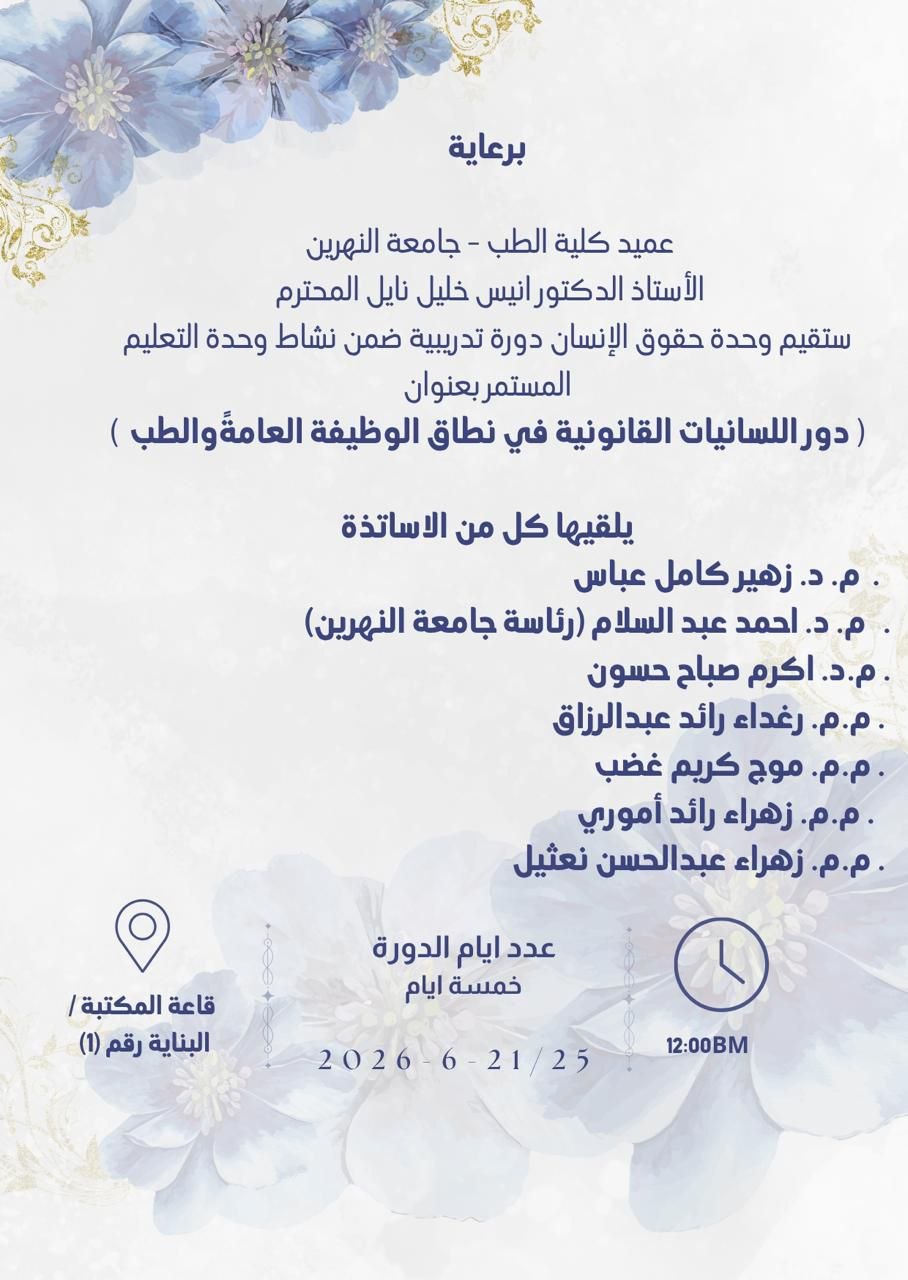
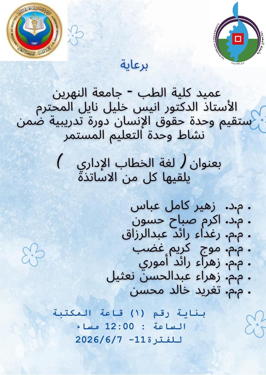
برعاية عميد كلية الطب – جامعة النهرين الأستاذ الدكتور أنيس خليل نايل تقيم وحدة حقوق الإنسان دورة تدريبية ضمن نشاطات وحدة التعليم المستمر بع...