

Master’s Thesis Defense at Al-Nahrain College of Medicine Investigates the Effects of Zinc Oxide Nanoparticles on the Liver Tissue of Mouse Fetuses
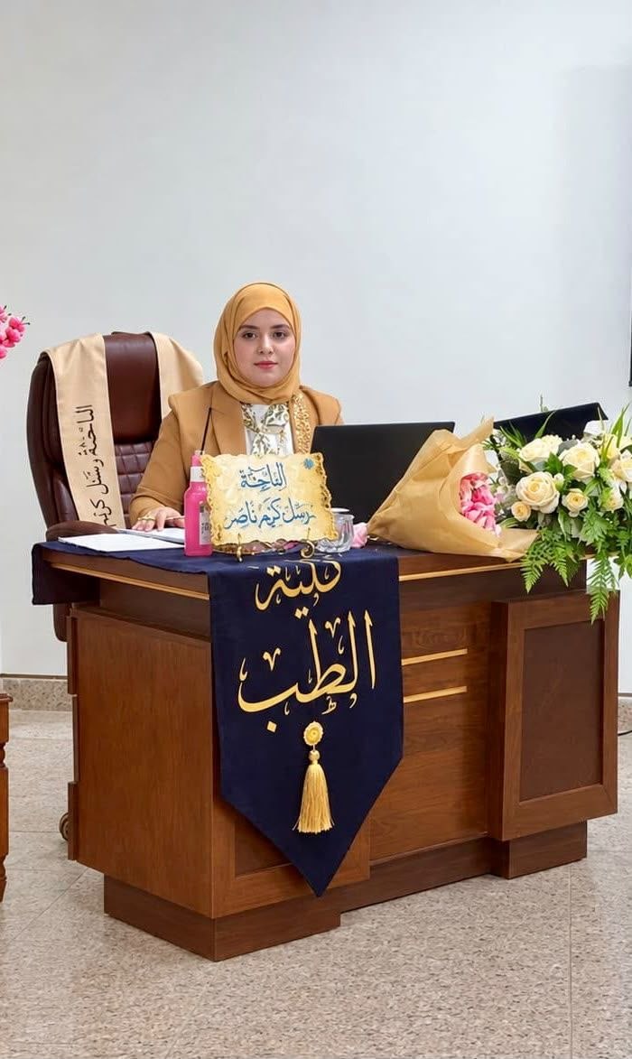
On Thursday, August 20, 2026, a Master’s thesis defense was held at the College of Medicine, Al-Nahrain University, Department of Physiology and Medical Physics, by Master’s student Rassel Kareem Nasser, entitled:
“Biophysical and Histological Assessment of Prenatal Zinc Oxide Nanoparticle Toxicity in Neonatal Mouse Liver Tissue via Tumor Necrosis Factor Profiling”
The thesis was supervised by Prof. Dr. Haider Abdul-Rasool Jaafar and Asst. Prof. Dr. Mazen Kamel Hamid.
Aim of the Study
The study aimed to investigate histological changes in the liver tissue of mouse fetuses following maternal exposure during pregnancy to chemically synthesized zinc oxide nanoparticles (ZnONPs) of a defined size and at different doses.
It also aimed to identify and characterize any structural alterations, cellular damage, or developmental abnormalities in fetal liver tissue.
Materials and Methods
In this study, zinc oxide nanoparticles (ZnONPs) were synthesized using a simple, low-cost chemical precipitation method. Their physicochemical characteristics, including particle size, morphology, and surface charge, were determined.
Forty adult female Swiss albino mice, weighing 20–25 g, were used in the study. The animals were provided by the Animal Care Unit at the College of Medicine, Al-Nahrain University. They were divided into five groups, with eight females in each group, and maintained under standard conditions in the College’s Animal Care Unit. The animals were kept under a 12-hour light/dark cycle, with standard rodent feed and water provided ad libitum.
The animals were subsequently divided into the following groups:
-
Group A (Control Group): Injected with distilled water.
-
Group B: Injected with zinc oxide nanoparticles at a dose of 5 mg/kg.
-
Group C: Injected with zinc oxide nanoparticles at a dose of 10 mg/kg.
-
Group D: Injected with zinc oxide nanoparticles at a dose of 20 mg/kg.
-
Group E: Injected with zinc oxide nanoparticles at a dose of 30 mg/kg.
Liver tissues obtained from neonatal mice were fixed in 10% formalin and processed into paraffin blocks for histological sectioning and subsequent morphometric, histological, and immunohistochemical assessments of TNF expression in neonatal mouse liver tissue.
The paraffin blocks were sectioned at a thickness of 4 µm and stained with hematoxylin and eosin (H&E). Immunohistochemical analysis was then performed to assess TNF protein expression using anti-TNF antibodies.
Results
The size of the synthesized zinc oxide nanoparticles (ZnONPs) was determined to be approximately 20–25 nm.
Maternal administration of zinc oxide nanoparticles during pregnancy resulted in a reduction in the thickness of hepatic cell plates and cords in fetal liver tissue, with shrinkage becoming more pronounced in animals treated with higher doses compared with those receiving lower doses and the control group. The differences were statistically significant (p < 0.001).
An increase in hepatocellular death was also observed, accompanied by severe necrosis of the hepatic tissue. Some cells appeared absent or disrupted, with pyknotic nuclei and vacuolated or degenerated cytoplasm.
These findings may indicate interactions or mechanisms of association between zinc oxide nanoparticles and red blood cells (RBCs). Immunohistochemical (IHC) examination revealed dark-brown staining produced by the DAB chromogen in the stained liver tissue samples.
Conclusions
The study concluded that zinc oxide nanoparticles (ZnONPs) appear to be relatively compatible with red blood cells at low concentrations, whereas at higher concentrations, they become more aggressive and induce greater damage to living cells.
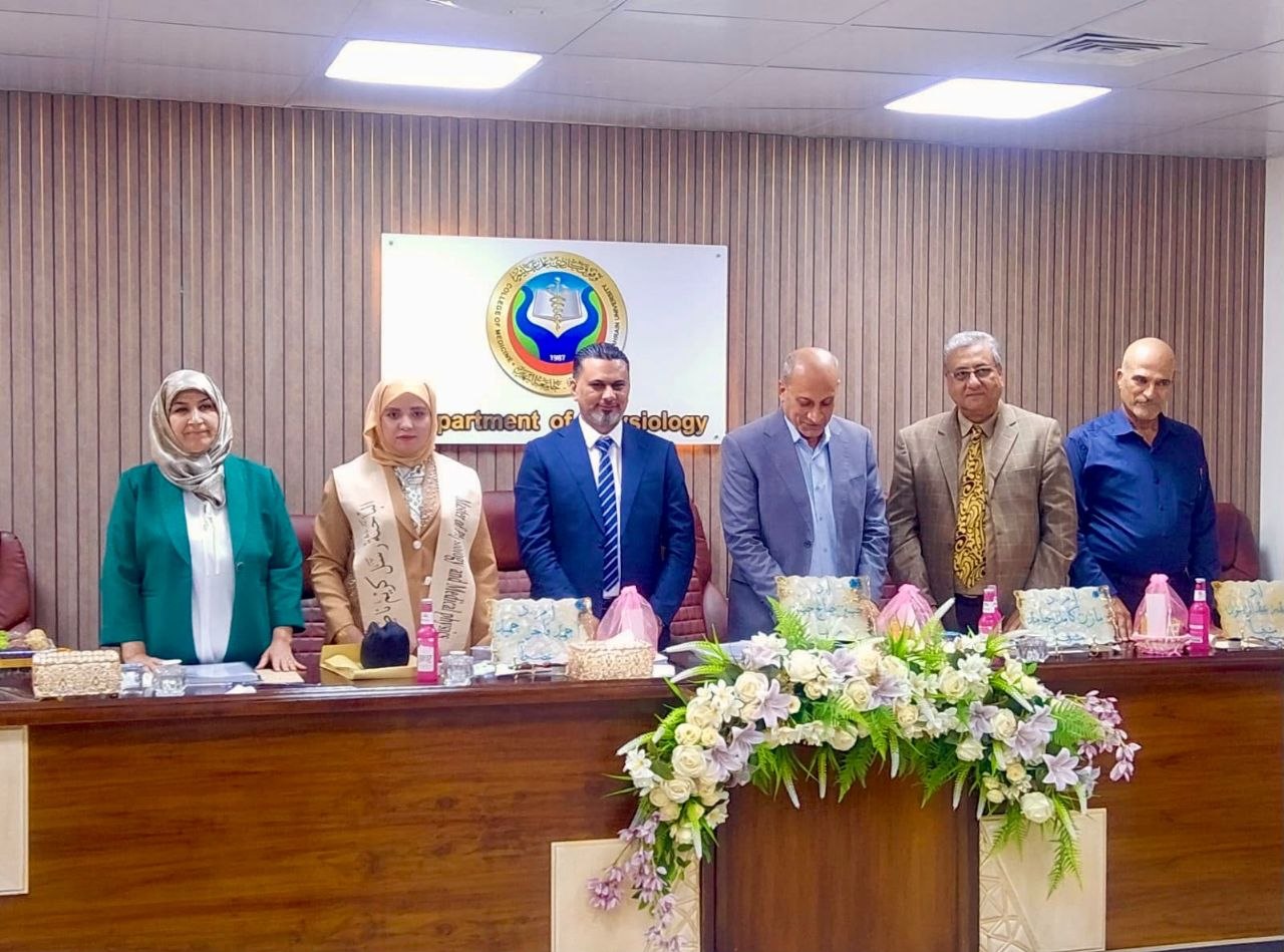
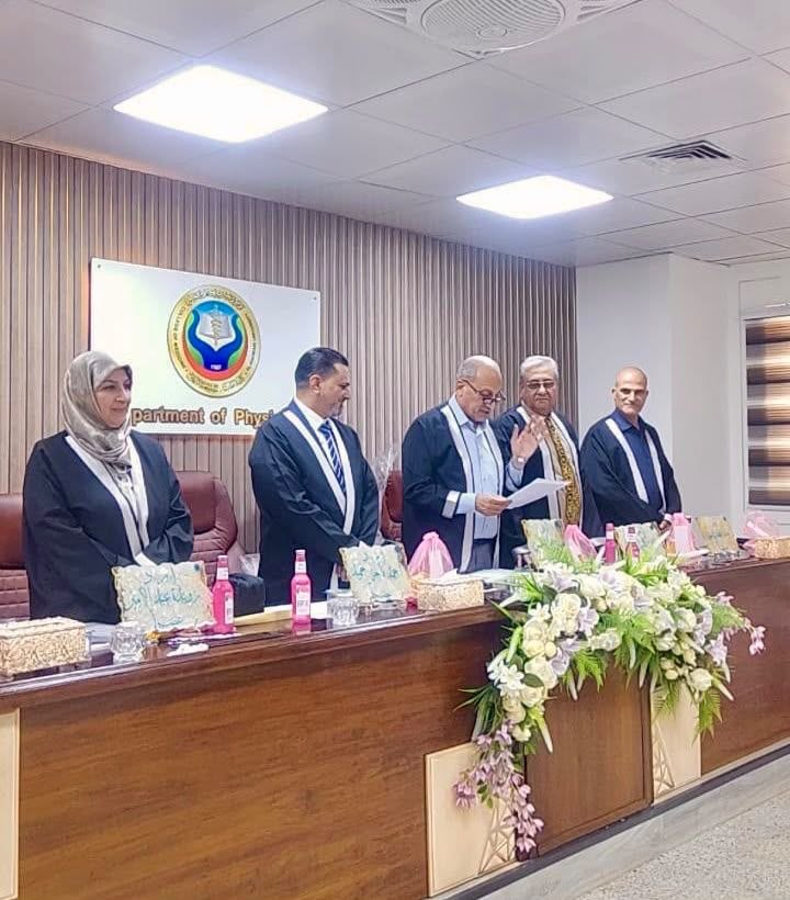
Thesis Defense Committee
The defense committee consisted of:
-
Prof. Dr. Hussein Saleh – Chair
-
Asst. Prof. Dr. Ruwaida Abdul-Amir – Member
-
Asst. Prof. Dr. Ahmed Fakher Hamid – Member
-
Asst. Prof. Dr. Mazen Kamel Hamid – Member and Supervisor
-
Prof. Dr. Haider Abdul-Rasool Jaafar – Member and Supervisor
The thesis was successfully defended and approved with an “Excellent” grade.
Media and Government Communication Division
College of Medicine – Al-Nahrain University