

Master’s Thesis Defense at Al-Nahrain College of Medicine Evaluates Silver Nanoparticles as an X-ray Contrast Agent in a Murine Model
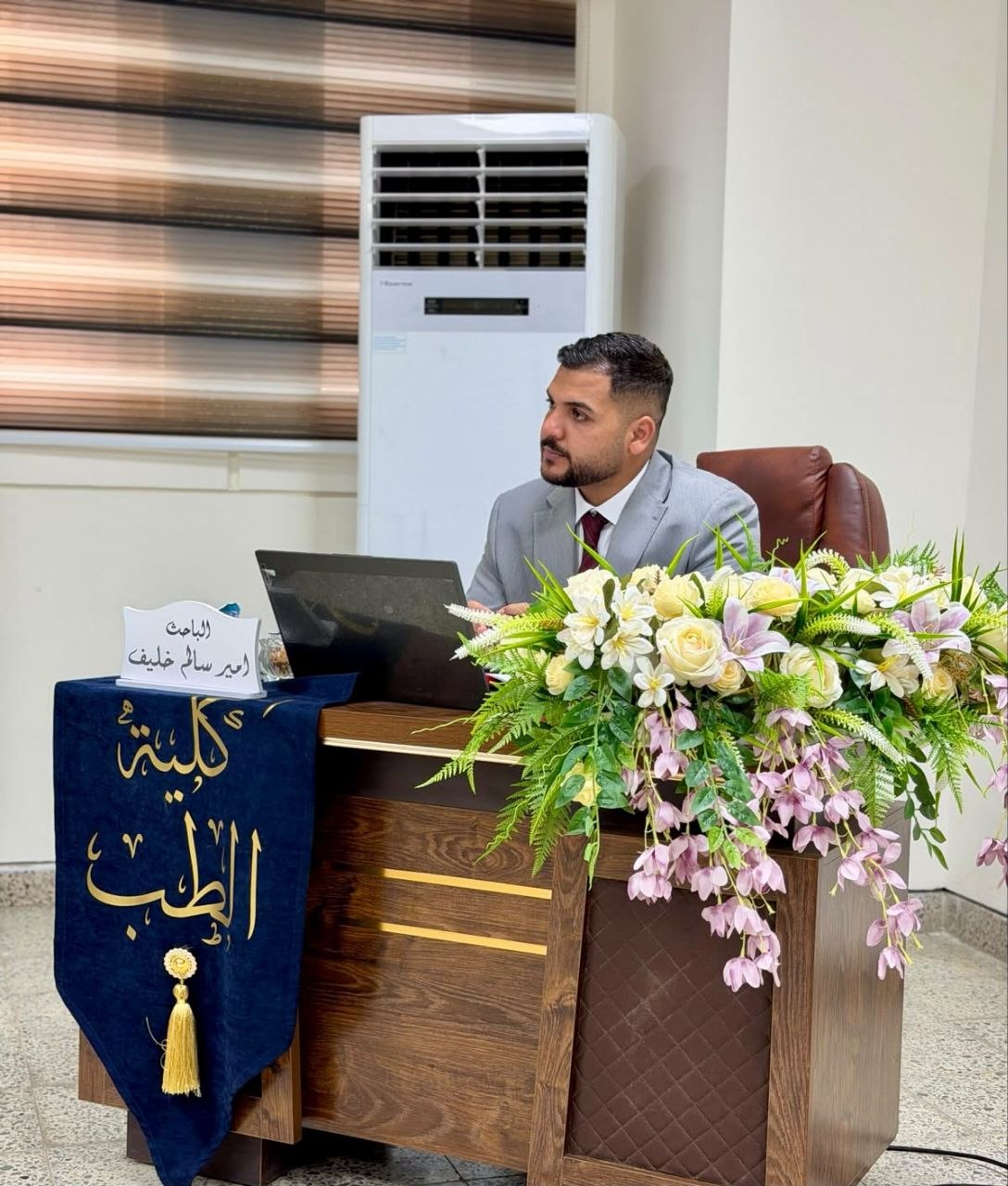
A Master’s thesis defense was held at the College of Medicine, Al-Nahrain University, Department of Physiology and Medical Physics, by Master’s student in Medical Physics Amir Salem Khalaf, entitled:
“In Vivo Evaluation of Silver Nanoparticles as an X- ray Contrast Agent in a Murine Model Comparison With an Iodine – Based Contrast Agent”
The thesis was supervised by Asst. Prof. Dr. Mazen Kamel Hamid and Lecturer Dr. Doaa Abdul-Hameed.
Aim of the Study
The study aimed to evaluate the performance of silver nanoparticles (AgNPs) administered intravenously as an in vivo X-ray contrast agent in mice, and to quantitatively compare the quality of the resulting images with that obtained using an iodine-based contrast agent, while deionized water was used as the control.
Materials and Methods
Healthy male mice, with an average body weight of approximately 20 g, received a single intravenous injection of 0.2 mL through the lateral tail vein.
The injected materials included chemically synthesized silver nanoparticle solutions at doses of 0.186, 0.25, 0.50, and 0.75 mg/kg, as well as silver nanoparticles at a concentration of 1.00 g/kg, an iodine-based contrast agent at a concentration of 0.30 mg/kg, and deionized water.
X-ray images were acquired at 1, 3, and 5 minutes following injection. Quantitative image analysis was performed on defined regions of interest under standardized imaging conditions.
The evaluated parameters included mean intensity, root mean square (RMS) contrast, Michelson contrast, entropy, and standard deviation.
Results
The group receiving silver nanoparticles at a dose of 0.75 mg/kg demonstrated the best overall image-quality performance, particularly 3 minutes after injection.
At this time point, this group showed the highest mean image intensity (84.56 units) and the highest entropy (7.31), exceeding the corresponding values observed in the iodine group.
Root mean square contrast and Michelson contrast were found to be useful complementary measures; however, their values were interpreted cautiously, as higher values may also reflect image noise, saturation, or heterogeneous distribution of the nanoparticles.
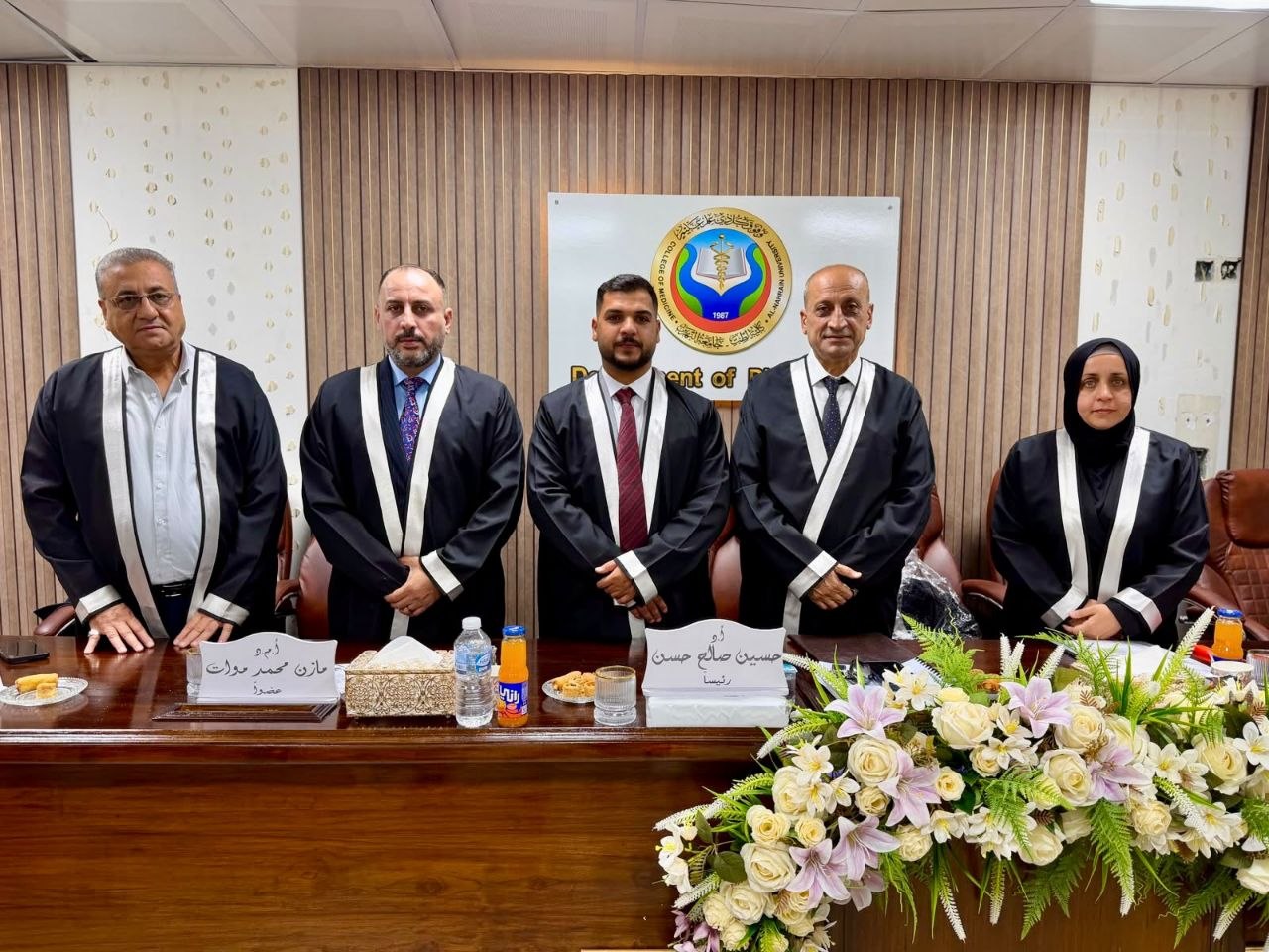
Thesis Defense Committee
The defense committee consisted of:
-
Prof. Dr. Hussein Saleh Hassan – Chair
-
Asst. Prof. Noor Kadhim – Member
-
Asst. Prof. Dr. Mazen Mohammed Muat – Member
-
Asst. Prof. Dr. Mazen Kamel Hamid – Member and Supervisor
-
Lecturer Dr. Doaa Abdul-Hameed – Member and Supervisor
The thesis was successfully defended and approved with an “Excellent” grade.
Media and Government Communication Division
College of Medicine – Al-Nahrain University